MATERIALS AND METHODS
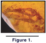 The
specimen, SMU 74976 (Figure 1), was
donated to the Shuler Museum of Paleontology at Southern Methodist University (SMU)
by William S. Lowe of Granbury, Texas, who discovered it in a commercial
shipment of amber containing plant and insect inclusions. The amber originated
in the Dominican Republic, but was purchased through a broker without precise
locality data. Dominican amber deposits are considered to be late Early to early
Middle Miocene in age, approximately 15-20 million years old (Iturralde-Vinent
and MacPhee 1996).
The
specimen, SMU 74976 (Figure 1), was
donated to the Shuler Museum of Paleontology at Southern Methodist University (SMU)
by William S. Lowe of Granbury, Texas, who discovered it in a commercial
shipment of amber containing plant and insect inclusions. The amber originated
in the Dominican Republic, but was purchased through a broker without precise
locality data. Dominican amber deposits are considered to be late Early to early
Middle Miocene in age, approximately 15-20 million years old (Iturralde-Vinent
and MacPhee 1996).
Preserved elements include a relatively complete
skull, portions of the first six cervical vertebrae, some soft tissue, and
limited squamation. The posterior-most vertebra is exposed to the polished amber
surface; the remaining portion of the fossil is completely encapsulated. Because
of the exposure of an articulated vertebra on the surface of the amber, we
assume the fossil was originally more complete (e.g., not preserved only as a
head and neck), but that the loss of an undetermined amount of the body occurred
between fossilization and curation. The amber is dark, approximately 1.5 cm x 1
cm x .85 cm, and contains gas bubbles and organic detritus. Semi-opacity of the
amber and optical distortion caused by its irregular, polished surface limits
microscopic examination.
CT scanning was performed at the University of
Texas High-Resolution X-ray CT Facility in Austin, Texas. The first scan used
3-slice-mode in two passes. Because of a computer error, the two passes did not
line up correctly, corrupting the three- dimensional reconstruction. However,
this scan provided enhanced detail on a slice-by-slice basis compared to the
second scan series. Settings for the first scan were: 80 kV, 0.2 mA, no filter,
air wedge, 160 per cent offset, slice thickness of 0.035 mm, standard outside
diameter (S.O.D.) 19 mm, 2696 views, two samples per view, inter-slice spacing
0.06 mm, field of reconstruction 4.5 mm, reconstruction offset 190,
reconstruction scale 7. Eight-bit export parameters were set to level 1600,
width 4095. The second scan was done using single-slice mode and thicker slices
to provide data adequate for 3D reconstruction. Settings used were: 80 kV, 0.2
mA, no filter, air wedge, no offset, slice thickness 0.068 mm, S.O.D. 22 mm,
1800 views, 2 samples per view, inter-slice spacing 0.06 mm, field of
reconstruction 4.5 mm, reconstruction offset 190, reconstruction scale 7.
Eight-bit export parameters were set to level 1500, width 3000.
The second scan resulted in 148 two-dimensional
images that were used to reconstruct pseudo-three-dimensional images using
Voxblast version 3.0 (Vaytek 2000).
Two-dimensional images were analyzed using Scion Image for Windows version Beta
3b (Scion 1998). A polygon surface
model was generated using the Voxblast iso-surface extraction function with a
threshold value of 120. The polygon surface model was then imported into
Lightwave version 6.5 (Newtek 2001)
to produce the final renderings. Two artifacts of the iso-surface extraction
algorithm are obvious. First, variability in the recorded density at the
bone-amber interface manifests as occasional non-reconstruction of any bone that
falls below the selected threshold value of 120, as in the void illustrated in Figure
5.4. Any artifacts in the iso-surface reconstruction that could affect
presence-absence or character state decisions (e.g., splenial, postfrontal) were
checked against the original two- dimensional data. Surface texturing manifested
as transverse raised ridges, most notably on curved surfaces, is due to sampling
density in the longitudinal axis and does not reflect the state of the bone
surface in the original specimen.
Descriptive nomenclature for osseous elements
follows Estes et al. (1988).
Institutional abbreviations are as listed in Leviton
et al. (1985). A complete set of digital data is on file at the Shuler
Museum of Paleontology at SMU and at the University of Texas High-Resolution
X-ray CT Facility.