DESCRIPTION OF THE SKULL (SMU 74976)
The head appears to have been partially de-fleshed
prior to encapsulation in the amber matrix (Figure
1). Skin is visible on the left side of the skull. Description of the scale
pattern, as observed under a dissecting microscope, follows Williams
et al. (1995). Taphonomic damage due to soft tissue decomposition adds
uncertainty to some of the scale counts. The rostral is broad. There are seven
supralabials to below the center of the eye. Three preoculars and three
suboculars are preserved, although it is unclear whether the preserved series is
an accurate reflection of the total number in life. The suboculars are in
contact with the supralabials. Four canthals are clearly discernable. A minimum
of 35 loreals are preserved with five loreal rows present. Ventrally, there are
two sublabial and three postmental scales (including sublabials) on each side of
the midline, for a total of six postmentals. The gular region, preserved as a
semi-transparent membrane, lacks gular folds.
 The
skull is 7.44 mm in length and 3.74 mm wide as measured across the jugals,
similar in size and proportion to the two other amber- preserved specimens (10.5
x 4.5 for AMNH DR-SH-1, ~8.5 x 4.5 for the NMBA specimen; de Queiroz et al.
1998). It is relatively complete, with damage restricted primarily to the right
lateral and posterodorsal regions.
The
skull is 7.44 mm in length and 3.74 mm wide as measured across the jugals,
similar in size and proportion to the two other amber- preserved specimens (10.5
x 4.5 for AMNH DR-SH-1, ~8.5 x 4.5 for the NMBA specimen; de Queiroz et al.
1998). It is relatively complete, with damage restricted primarily to the right
lateral and posterodorsal regions.
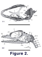
The skull has a T-shaped, unpaired premaxilla with
a narrow internarial bar that extends posteriorly to about the third or fourth
maxillary tooth position (Figure 2, Figure
3, Figure 4). Lateral premaxillary
processes form the anterior floor of the external nares. Two sub-conical
premaxillary teeth are preserved, one each in the lateral position of each side,
with room for a total of five or six tooth positions. Articulation with the
maxilla is oblique and loose. Septomaxillae and nasals are not preserved.
 The
maxillae are damaged along their dorsomedial margins. Maxillary palatine
processes are well developed, protruding medially at about the seventh maxillary
tooth position. The maxillae are depressed in lateral view to form the posterior
floor of the external nares. Thirteen or 14 tooth positions are preserved in
each maxilla, the tooth row extending posteriorly beyond the ectopterygoid
contact. The left prefrontal is nearly complete, the right is badly damaged. The
maxillary-prefrontal sutures are indistinct, but the junction presents a
continuous surface. The prefrontals probably did not contact the nasal, as the
apparently complete anterolateral process of the frontal appears to exclude this
contact.
The
maxillae are damaged along their dorsomedial margins. Maxillary palatine
processes are well developed, protruding medially at about the seventh maxillary
tooth position. The maxillae are depressed in lateral view to form the posterior
floor of the external nares. Thirteen or 14 tooth positions are preserved in
each maxilla, the tooth row extending posteriorly beyond the ectopterygoid
contact. The left prefrontal is nearly complete, the right is badly damaged. The
maxillary-prefrontal sutures are indistinct, but the junction presents a
continuous surface. The prefrontals probably did not contact the nasal, as the
apparently complete anterolateral process of the frontal appears to exclude this
contact.
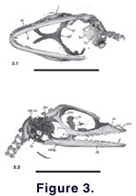
The anterior roof table is formed by the frontal,
which is relatively complete. Diverging anterior processes extend
anterolaterally to contact the prefrontals and medially exhibit a shelf-like
facet for articulation with the nasals. There is no evidence of an anterior
medial process as seen in A. carolinensis (Stimie
1966), but instead the frontal forms a v-shaped cleft. The frontal is
strongly constricted between the orbits, forming their complete dorsal margin.
Discernable parietal remnants occur only at the lateral frontal-parietal suture;
however, an inverted cone-like notch is preserved at the midline of the suture (Fig.
3.1, Fig. 4.2) and may represent the
anterior wall of the pineal foramen. Postfrontals are not present as in the NMBA
specimen. Although a left postfrontal is reported on the AMNH specimen,
examination of the stereo-radiograph in de
Queiroz et al. (1998) illustrates the element in a damaged portion of the
skull and suggests it may as likely represent a fragment of the frontal.
 The
left lacrimal is present, forming the anteroventral margin of the orbit, and
meeting the anterior margin of the jugal posteroventrally. The anterior terminus
of the jugal is lateral to the posterior maxillary tooth position. The jugal
forms the greater portion of the ventral and posterior orbital margin, and
overlies the lateral face of the postorbital posteriorly. There is no contact of
the jugal with the squamosal. The tri-radiate postorbitals are complete; the two
anterior processes complete the posterodorsal orbital margin, while the third
extends posteriorly to contact the squamosal, although this junction is not
clearly defined. The rod-like squamosal lies lateral to the preserved left
supratemporal and does not appear to have had contact with the parietal. It
terminates posteriorly in a hook-like, ventrally directed process that extends
into the tympanic recess of the quadrate.
The
left lacrimal is present, forming the anteroventral margin of the orbit, and
meeting the anterior margin of the jugal posteroventrally. The anterior terminus
of the jugal is lateral to the posterior maxillary tooth position. The jugal
forms the greater portion of the ventral and posterior orbital margin, and
overlies the lateral face of the postorbital posteriorly. There is no contact of
the jugal with the squamosal. The tri-radiate postorbitals are complete; the two
anterior processes complete the posterodorsal orbital margin, while the third
extends posteriorly to contact the squamosal, although this junction is not
clearly defined. The rod-like squamosal lies lateral to the preserved left
supratemporal and does not appear to have had contact with the parietal. It
terminates posteriorly in a hook-like, ventrally directed process that extends
into the tympanic recess of the quadrate.
Only the left quadrate is preserved. It is
anteroventrally oriented and the main shaft is long and narrow (4:1
length/width). The tympanic rim is well developed, extending from the squamosal
to the posterolateral shelf that lies dorsal to the articular facet of the
mandibular fossa, forming a shallow concavity.
Of the palatal bones, the left and right
ectopterygoids and left pterygoid are present. The left ectopteryogoid-pterygoid
contact is unclear. The ectopterygoid contacts the jugal near the posterior
terminus of the maxilla. Pterygoid teeth are not present, and there is no
contact of the pterygoid (or ectopterygoid) with the lacrimal.
There is significant damage to the braincase and
little of this region could be resolved. Of the basicranial elements, the
parabasisphenoid is absent and the basioccipital is fragmentary. The left
prootic and opisthotic are present, exposing the osseous labyrinth medially.
Within the osseous labyrinth, the posterior semi-circular canals, which do not
exhibit prominent ridges on their surfaces, are visible. Nothing could be
discerned concerning fusion or suturing of contacts. Within the fenestra ovalis
there is the remnant of a ring-like ossifcation that may be the stapedial
footplate.
 The
mandibles are relatively well preserved (Figure
5). The dentaries each bear 17-18 tooth positions. The anterior teeth are
sub-conical, with tricuspid tooth morphology appearing at about the tenth tooth
position. There is no sculpting of the dentaries. However, the left dentary
displays a laterally and medially directed ossified protuberance that may
represent a pathology. As many as six mental foramina are present on the lateral
surface of each dentary. Meckel's groove is enclosed to the posterior tooth
position. The posteromedial end of the dentary is divided into dorsal and
ventral processes; the dorsal process terminates against the anterior process of
the coronoid while the ventral process tapers posteriorly, terminating ventral
to the apex of the coronoid. Laterally, the dentary meets the surangular in a
poorly defined suture.
The
mandibles are relatively well preserved (Figure
5). The dentaries each bear 17-18 tooth positions. The anterior teeth are
sub-conical, with tricuspid tooth morphology appearing at about the tenth tooth
position. There is no sculpting of the dentaries. However, the left dentary
displays a laterally and medially directed ossified protuberance that may
represent a pathology. As many as six mental foramina are present on the lateral
surface of each dentary. Meckel's groove is enclosed to the posterior tooth
position. The posteromedial end of the dentary is divided into dorsal and
ventral processes; the dorsal process terminates against the anterior process of
the coronoid while the ventral process tapers posteriorly, terminating ventral
to the apex of the coronoid. Laterally, the dentary meets the surangular in a
poorly defined suture.
The ventral extension of the labial process of the
coronoid is unclear. The anteromedial process extends ventrally to terminate
without a posterior projection, and the posteromedial process contacts the
articular. There is no splenial. It is unclear if the angular is present. The
surangular forms the dorsal margin of the lower jaw posterior to the coronoid
process. There is a well-developed medial angular process of the articular.