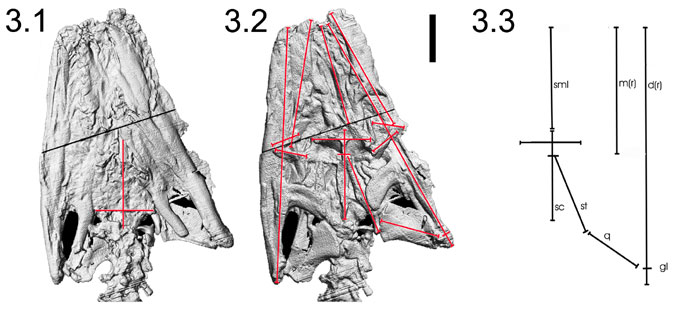
Figure 3. Three-dimensional isosurface models derived from CT data and line segment stand-ins to test alignment and displacement of cranial elements of Pachyrhachis problematicus (HUJ-PAL 3659). 3.1, Ventral view with lines indicating sagittal plane traced from the median basioccipital to the parasphenoid rostrum and transverse planes indicating lateral limits of exoccipitals. 3.2, Dorsal view with lines indicating sagittal and transverse planes traced on parietal, and line segments to act as stand-ins for major elements and landmarks. 3.3, Line segment stand-ins, rotated and adjusted to remove translational and rotational distortion of major elements. Abbreviations: d, dentary; gl, glenoid; m, maxilla; pob, postorbital; q, quadrate; sc, sagittal crest; sml, sagittal mid-line; st, supratemporal. Scale bar equals one centimeter.