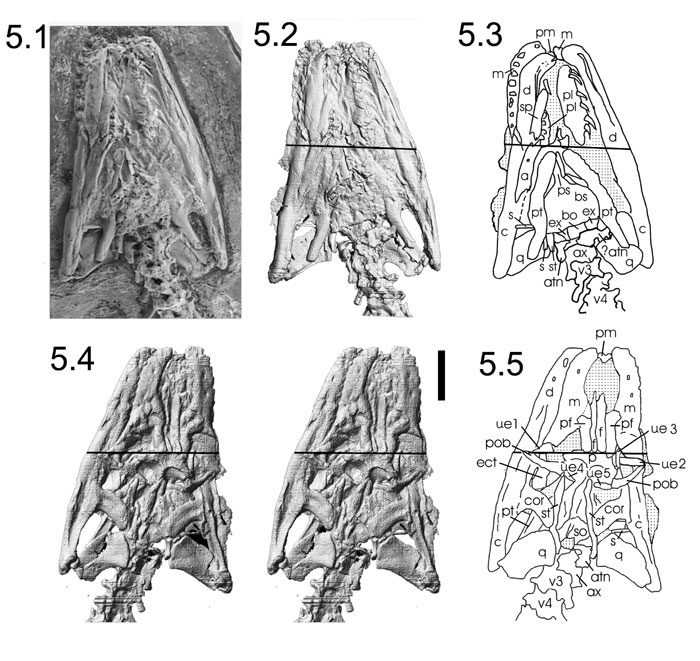
Figure 5. Photograph and computer reconstructions derived from CT data, and interpretive line drawings of Pachyrhachis problematicus (HUJ-PAL 3659). 5.1, light photograph of ventral surface as currently conserved; 5.2, computer reconstructed isosurface model derived from CT data. Note resolution and fidelity of surface reconstruction as compared with light photograph. 5.3, interpretive drawing of ventral surface; 5.4, stereopair of dorsal surface from computer reconstructed surface model derived from CT data; 5.5, interpretive drawing of dorsal surface. Abbreviations: a, angular; atn, atlas neural arch; ax, axis vertebra; bs, basisphenoid; bo, basioccipital; c, compound bone; cor, coronoid; d, dentary; ect, ectopterygoid; ex, exoccipital; f, frontal; m, maxilla; p, parietal; pm, premaxilla; pob, postorbital; pf, prefrontal; pl, palatine; pt, pterygoid; q, quadrate; s, stapes; so, supraoccipital; sp, splenial; st, supratemporal, ue1-5; unidentified elements; v3-v4, 3rd and 4th cervical vertebra. (See also Appendix 2, Appendix 3.) Scale bar equals one centimeter.