|
|
APPENDIX 4. DESCRIPTIVE TAXONOMY CUT-L-EEH
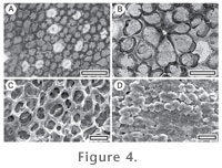
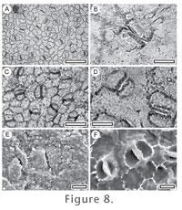
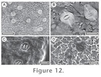
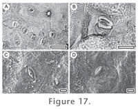
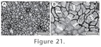
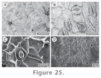
Referred specimens and occurrence: SL3179, BL-01; SL2626, BL-07; SL2258, BL-15; SL1174, GL-01; SL2674, GL-04; SL2137, GL-08; SL3286, Mata-03; SL1550, Sthd-002; SL1577, Sthd-004; SL1747, Sthd-016; SL1727, Sthd-017; SL1774, Sthd-022; SL1793, Sthd-029; SL1960, Sthd-054; SL1905, Sthd-074; SL2020, Sthd-087; SL1850, Sthd-088; SL1813, Sthd-100. DescriptionStomatal complexes. Stomatal distribution over leaf surfaces unknown; stomatal complexes in clear areoles; isolated; randomly oriented; paracytic; outline rounded, but irregular; length 16–22 m (medium); stomatal size range bimodal, with distinct 'giant stomatal complexes' present. Subsidiary cell periclinal cuticle thinner than over normal epidermal cells, smooth, unornamented. Guard cells overarched by subsidiary cells; Stomatal pore slit-like; cuticular scales narrow; not clear. Epidermal Cells. Epidermal cell flanges clearly visible using TLM; cells over fine venation distinguished as 'venal' (elongated); normal epidermal cells isodiametric; walls straight; unbuttressed. Indumentum. Outer surface smooth; unornamented; with scars of trichome bases and papillose. Papillae present over all normal epidermal cells, and some, but not all, venal cells; 1–4 papillae per cell; formed by the projection of a discrete region within the boundaries of the epidermal cell; smooth. Trichomes sparse; restricted to regions over veins; deciduous (and therefore trichome type unknown); inserted between epidermal cells; diameter generally smaller than a normal epidermal cell; epidermal cells around trichome base modified with thickened poral rim and radial walls, radially elongated as distinct foot cells (5–6); trichome bases sometimes paired. Distinguishing features. The multi-headed papillae make CUT-L-EEH distinct from all the other Lauraceae taxa. Identification. Not similar to any species illustrated by Christophel and Rowett (1996). No extant species of Australasian Lauraceae appear to have multi-lobed papillae.
CUT-L-DFI (Cryptocarya sp.)
Referred specimens and occurrence: SL2530, BL-05; SL2452, BL-32; SL2656, GL-04; SL2912, GL-21; SL2955, GL-25; SL3001, GL-27; SL0065, Mata-18; SL1561, Sthd-004; SL1735, Sthd-017; SL1883, Sthd-073; SL1596, Sthd-108; SL2079, Sthd-113. DescriptionStomatal complexes. Stomatal distribution over leaf surfaces hypostomatic; stomatal complexes in clear areoles; isolated; randomly oriented; paracytic; outline rounded, but irregular; length 10–15 m (small); stomatal size range unimodal with a small range. Subsidiary cell periclinal cuticle thinner than over normal epidermal cells; smooth; unornamented. Guard cells overarched by subsidiary cells. Stomatal pore slit-like; cuticular scales 'butterfly-like', about two-thirds the length of the subsidiary cells. Epidermal Cells. Epidermal cell flanges clearly visible using TLM; cells over fine venation distinguished as 'venal' (elongated); normal epidermal cells isodiametric; walls sinuous, or curved or wavy; unbuttressed. Indumentum. Outer surface smooth; unornamented; with scars of trichome bases and papillose. Papillae present over all epidermal cells; 1 papilla per cell; formed by the entire outer surface of the epidermal cell projecting upwards; smooth. Trichomes common; scattered over venal and non-venal regions; deciduous (and therefore trichome type unknown); inserted between epidermal cells; diameter similar in size to a normal epidermal cell; epidermal cells around trichome base (6–9) modified with thickened poral rim and radial walls, forming a distinct ring of isodiametric foot cells. Non-stomatal surface. Epidermal cells isodiametric; polygonal; cells over veins not distinguished. Simple trichome bases present. Distinguishing features. The epidermal cells which are entirely raised up as papillae make CUT-L-DFI distinct from all the other Lauraceae taxa. Identification. The distinct cuticular scales suggest Cryptocarya, although the broadly papillate nature of the epidermal cells is unlike any extant Australasian species (Christophel and Rowett (1996)). It is probably an extinct species of Cryptocarya.
CUT-L-DEJ (Litsea sp.)
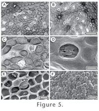
Referred specimens and occurrence: SL2220, GL-12. DescriptionStomatal complexes. Stomatal distribution over leaf surfaces unknown; stomatal complexes evenly spread; isolated; randomly oriented; paracytic; outline typically broader than long; length 25–30 m (medium); stomatal size range unimodal with a small range. Subsidiary cell periclinal cuticle not distinct in thickness from normal epidermal cells; smooth; unornamented. Guard cells overarched by subsidiary cells; Stomatal pore plugged; cuticular scales narrow, but massively thickened; full length of subsidiary cells (appearing very distinct under TLM). Epidermal Cells. Epidermal cell flanges not clearly visible under TLM; cells over major veins distinguished as 'venal' (elongated); normal epidermal cells unclear; walls unclear; unbuttressed. Indumentum. Outer surface smooth; unornamented; papillose. Papillae present over all epidermal cells; 1 papilla per cell; formed by the projection of a discrete region within the boundaries of the epidermal cell; smooth. Distinguishing features. CUT-L-DEJ has discrete papillae within the bounds of the epidermal cell outline. This is similar to CUT-L-ECI but cuticle thickness/robustness is much less. Identification. The broad, well-defined papillae are similar to four species of Litsea (L. bennettii B. Hyland, L. breviumbellata C.K. Allen, L. connorsii B.Hyland, L. leefeana (F. Muell.) Merr.) as illustrated by Christophel and Rowett (1996) and indicate identity with that genus.
CUT-L-ECI
Referred specimens and occurrence: SL1763, Sthd-011; SL1610, Sthd-108. Description Stomatal complexes. Stomatal distribution over leaf surfaces unknown; stomatal complexes evenly spread; isolated; randomly oriented; paracytic; outline typically broader than long; length c. 35 m (medium); stomatal size range unimodal with a small range. Subsidiary cell periclinal cuticle not distinct in thickness from normal epidermal cells; smooth; unornamented. Guard cells overarched by subsidiary cells; Stomatal pore plugged; cuticular scales narrow; not clear. Epidermal Cells. Epidermal cell flanges not clearly visible under TLM; cells over veins not distinguished by shape; normal epidermal cells unclear; walls unclear; unbuttressed. Indumentum. Outer surface smooth; unornamented; papillose. Papillae present over all epidermal cells; (probably) just 1 papilla per cell; formed by the projection of a discrete region within the boundaries of the epidermal cell; smooth. Distinguishing features. CUT-L-ECI has discrete papillae within the bounds of the epidermal cell outline. This is similar to CUT-L-DEJ but cuticle thickness/robustness is much greater. Identification. The thick-walled papillae are similar to Cryptocarya mackinnoniana F.Muell. and Endiandra palmerstonii (F.M.Bailey) C.T. White & W.D.Francis as illustrated by Christophel and Rowett (1996), but nothing else about the fossil is convincingly similar. Its generic identity remains unclear.
CUT-L-DEC
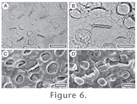
Referred specimens and occurrence: SL1278, BL-05; SL3084, BL-28; SL1311, BL-32; SL1181, GL-01; SL2657, GL-04; SL2099, GL-07; SL2134, GL-08; SL2162, GL-09; SL2215, GL-12; SL2826, GL-16; SL2841, GL-18; SL2892, GL-20; SL3103, GL-24; SL2959, GL-25; SL2980, GL-26; SL2994, GL-27; SL2546, Mata-23; SL1827, Sthd-095. Description Stomatal complexes. Stomatal distribution over leaf surfaces hypostomatic; stomatal complexes in clear areoles; isolated; randomly oriented; paracytic; outline typically broader than long; length 25–30 m (medium); stomatal size range unimodal with a small range. Subsidiary cell periclinal cuticle not distinct in thickness from normal epidermal cells; granular; unornamented. Guard cells overarched by subsidiary cells; Stomatal pore massively thickened and with prominent T-piece thickenings at polar ends; cuticular scales narrow, but massively-thickened; full length of subsidiary cells (appearing very distinct under TLM). Epidermal Cells. Epidermal cell flanges clearly visible using TLM; cells over major veins distinguished as 'venal' (isodiametric); normal epidermal cells highly variable from isodiametric to elongate; walls sinuous, or curved or wavy; unbuttressed to buttressed. Indumentum. Outer surface coarsely granular; unornamented; with scars of trichome bases. Trichomes sparse; distribution not clear; deciduous (and therefore trichome type unknown); inserted between epidermal cells. Non-stomatal surface. Epidermal cells isodiametric; polygonal; cells over veins not distinguished. Distinguishing features. CUT-L-DEC is immediately recognisable based on its sinuous epidermal cell walls, the massive thickening around the stomatal; pores, and granular texture. Identification. The massively thickened stomatal aperture region is quite unlike anything illustrated by Christophel and Rowett (1996) and this fossil probably belongs to an extinct (at least locally) genus.
CUT-L-DCA
Referred specimens and occurrence: SL0232, BL-04; SB1284, BL-33; SL2954, GL-25; SL2017, Sthd-010. Description Stomatal complexes. Stomatal distribution over leaf surfaces unknown; stomatal complexes in clear areoles; isolated; randomly oriented; paracytic; outline typically broader than long; length 18–21 m (medium); stomatal size range unimodal with a small range. Subsidiary cell periclinal cuticle thinner than over normal epidermal cells; smooth; unornamented. Guard cells overarched by subsidiary cells; Stomatal pore slit-like; cuticular scales narrow. Epidermal Cells. Epidermal cell flanges clearly visible using TLM; cells over fine venation distinguished as 'venal' (elongated); normal epidermal cells isodiametric; walls sinuous; relatively thin, unbuttressed. Indumentum. Outer surface smooth; unornamented; with scars of trichome bases;. Trichomes common; restricted to regions over veins; deciduous (and therefore trichome type unknown); inserted between epidermal cells; diameter similar in size to a normal epidermal cell; epidermal cells around trichome base (5–8) modified with thickened poral rim and radial walls, radially elongated as distinct foot cells. Distinguishing features. CUT-L-DCA is recognised by its thin, sinuous epidermal cell walls. Identification. None of the species illustrated by Christophel and Rowett (1996) are particularly similar.
CUT-L-EHE
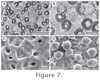
Referred specimens and occurrence: SL0308, BL-01; SB1340, BL-32. Description Stomatal complexes. Stomatal distribution over leaf surfaces hypostomatic; stomatal complexes in clear areoles; isolated; randomly oriented; paracytic; outline circular; length 15–23 m (medium); stomatal size range unimodal with a small range. Subsidiary cell periclinal cuticle thinner than over normal epidermal cells; smooth; unornamented. Guard cells overarched by subsidiary cells; Stomatal pore slit-like; cuticular scales 'butterfly-like'; about one-third the length of the subsidiary cells. Epidermal Cells. Epidermal cell flanges clearly visible using TLM; cells over fine venation distinguished as 'venal' (elongated); normal epidermal cells isodiametric; walls sinuous, or straight; slightly buttressed. Indumentum. Outer surface smooth; unornamented; with scars of trichome bases. Trichomes sparse; distribution not clear; deciduous (and therefore trichome type unknown); inserted between epidermal cells; diameter generally smaller than a normal epidermal cell; epidermal cells around trichome base (seven) modified with thickened poral rim and radial walls, unmodified in shape. Non-stomatal surface. Epidermal cells highly variable from isodiametric to elongate; walls slightly wavy; cells over veins distinguished by being longer and staining much darker than normal epidermal cells. Distinguishing features. Recognisable by the very rounded outline of the stomatal complexes in combination with slightly wavy epidermal cell walls. Identification. None of the species illustrated by Christophel and Rowett (1996) is particularly similar.
CUT-L-EEB
Referred specimens and occurrence: SL2163, GL-09; SL1786, Sthd-029; SL2027, Sthd-087. Description Stomatal complexes. Stomatal distribution over leaf surfaces unknown; stomatal complexes in clear areoles; isolated; randomly oriented; paracytic; outline rounded, but irregular; length 16–21 m (medium); there often appears to be a pair of epidermal cells, one on either side of the stomatal complex, which are probably part of the complex in the broader sense; stomatal size range unimodal with a small range. Subsidiary cell periclinal cuticle not distinct in thickness from normal epidermal cells; smooth; unornamented. Guard cells overarched by subsidiary cells; Stomatal pore slit-like; cuticular scales narrow; not clear. Epidermal Cells. Epidermal cell flanges clearly visible using TLM; cells over fine venation distinguished as 'venal' (elongated); normal epidermal cells isodiametric; walls sinuous; buttressed. Indumentum. Outer surface smooth; unornamented; glabrous. Distinguishing features. Recognisable by the very rounded outline of the stomatal complexes in combination with sinuous and buttressed epidermal cell walls and also the pair of epidermal cells on either side of the stomatal complex. Identification. The distinctive pair of epidermal cells on either side of most stomatal complexes makes this taxon distinct to any of the species illustrated by Christophel and Rowett (1996).
CUT-L-EHD
Referred specimens and occurrence: SL0102, BL-08; SL2534, BL-33; SL2821, GL-16; SL2074, Sthd-113. DescriptionStomatal complexes. Stomatal distribution over leaf surfaces unknown; stomatal complexes evenly spread; isolated; randomly oriented; paracytic; outline typically broader than long; length 20–27 m (medium); stomatal size range bimodal, with distinct 'giant stomatal complexes' present. Subsidiary cell periclinal cuticle thinner than over normal epidermal cells; smooth; unornamented. Guard cells overarched by subsidiary cells; Stomatal pore slit-like; cuticular scales double; very small. Epidermal Cells. Epidermal cell flanges clearly visible using TLM; cells over major veins distinguished as 'venal' (elongated); normal epidermal cells isodiametric; walls sinuous, or curved or wavy; buttressed. Indumentum. Outer surface smooth; unornamented; glabrous, or with scars of trichome bases. Trichomes sparse; deciduous (and therefore trichome type unknown); inserted between epidermal cells; epidermal cells around trichome base (3–9) modified with massively thickened poral rim, unmodified in shape. Distinguishing features. Distinguished by the sinuous and buttressed epidermal cells in combination with a stomatal complex which has very thin cuticle over the subsidiary cells. Identification. The sinuous and buttressed abaxial epidermal cells are found today in Beilschmiedia, Cryptocarya and Endiandra (Christophel and Rowett 1996). Not similar to any species illustrated by Christophel and Rowett (1996).
CUT-L-FJD (Beilschmiedia sp.)
DescriptionStomatal complexes. Stomatal distribution over leaf surfaces unknown; stomatal complexes in clear areoles; isolated; randomly oriented; paracytic; outline rounded, but irregular; length 8–10 m (small); development type often appear to be epidermal cells on either side of the stomatal complexes which are probably part of the complex in the broader sense; stomatal size range unimodal with a small range. Subsidiary cell periclinal cuticle not distinct in thickness from normal epidermal cells; granular; unornamented. Guard cells overarched by subsidiary cells; Stomatal pore slit-like; cuticular scales broad; full length of subsidiary cells (appearing very distinct under TLM). Epidermal Cells. Epidermal cell flanges clearly visible using TLM; cells over major veins distinguished as 'venal' (elongated); normal epidermal cells highly variable from isodiametric to elongate; walls curved or wavy; buttressed. Indumentum. Outer surface smooth; unornamented; with scars of trichome bases. Trichomes sparse; restricted to regions over veins; deciduous (and therefore trichome type unknown); inserted between epidermal cells; diameter generally smaller than a normal epidermal cell; epidermal cells around trichome base (6–7) modified with massively thickened poral rim, radially elongated as distinct foot cells. Distinguishing features. Distinguished by the buttressed (but not sinuous) epidermal cell walls in combination with broad scales.
CUT-L-DFA and MANU-3 (Cryptocarya sp.) Identification. The buttressed epidermal cells and the broadly thickened subsidiary cells indicate this taxon is likely Beilschmiedia (cf. B. brunnea B. Hyland illustrated by Christophel and Rowett 1996). MANU-3
Referred specimen and locality: SL1494, GL-01. Note. The mummified specimens found in this study fit the description of leaf parataxon MANU-3 described in Pole (1993a) reasonably well. There may be subtle differences in leaf shape, and much larger leaves were typical at the reference locality. A larger collection from the St Bathans Paleovalley may eventually show a distinct but probably closely related taxon. CUT-L-DFA
Referred specimens and occurrence: SL1349, BL-05; SL0095, BL-08; SL2272, BL-16; SL2115, GL-07; SL2132, GL-08; SL2174, GL-09; SL2193, GL-10; SL2915, GL-21; SL2917, GL-22; SL2933, GL-23; SL2948, GL-25; SL2987, GL-27; SL3255, GL-31; SL3033, GL-32. DescriptionStomatal complexes. Stomatal distribution over leaf surfaces hypostomatic; stomatal complexes in clear areoles; isolated; randomly oriented; paracytic; outline polygonal; length 18–30 m (medium); stomatal size range unimodal with a small range. Subsidiary cell periclinal cuticle not distinct in thickness from normal epidermal cells; smooth; unornamented. Guard cells overarched by subsidiary cells; Stomatal pore slit-like; cuticular scales 'butterfly-like'; full length of subsidiary cells (appearing very distinct under TLM). Epidermal Cells. Epidermal cell flanges clearly visible using TLM; cells over fine venation distinguished as 'venal' (elongated); normal epidermal cells highly variable from isodiametric to elongate; walls wavy; unbuttressed. Indumentum. Outer surface smooth; unornamented; glabrous. Non-stomatal surface. Epidermal cells isodiametric; polygonal; cells over veins not distinguished. Simple trichome bases present. Distinguishing features. CUT-L-DFA has small-moderately sized "butterfly" shaped scales in combination with smooth and wavy-walled epidermal cells. The adaxial epidermis is very distinctive in having venal epidermal cells which stain much darker than the normal epidermal cells. Identification. The small stomatal complexes with broad scales are broadly similar to some species of Cryptocarya (e.g. C. vulgaris B.Hyland) as illustrated by Christophel and Rowett (1996), and probably belong in that genus.
CUT-L-DGA (Cryptocarya sp.)
Referred specimens and occurrence: SL0303, BL-01; SL0245, BL-04; SL2632, BL-15; SL2267, BL-16; SL2285, BL-18; SL2305, BL-21; SL1310, BL-32; SL2679, GL-04; SL2117, GL-07; SL2153, GL-09; SL2181, GL-10; SL2195, GL-11; SL2212, GL-12; SL2845, GL-18. DescriptionStomatal complexes. Stomatal distribution over leaf surfaces hypostomatic; stomatal complexes in clear areoles; isolated; randomly oriented; paracytic; outline rounded, but irregular; length 12–20 m (medium); stomatal size range unimodal with a small range. Subsidiary cell periclinal cuticle thicker than over normal epidermal cells; smooth; unornamented. Guard cells overarched by subsidiary cells; Stomatal pore slit-like; cuticular scales 'butterfly-like'; full length of subsidiary cells (appearing very distinct under TLM). Epidermal Cells. Epidermal cell flanges clearly visible using TLM; cells over fine venation distinguished as 'venal' (elongated); normal epidermal cells highly variable from isodiametric to elongate; walls curved or wavy; unbuttressed. Indumentum. Outer surface smooth; unornamented; with scars of trichome bases. Trichomes sparse; restricted to regions over veins; deciduous (and therefore trichome type unknown); inserted between epidermal cells; diameter generally larger than a normal epidermal cell; epidermal cells around trichome base (5–7) unthickened, unmodified in shape. Non-stomatal surface. Epidermal cells isodiametric, unbuttressed; cells over veins somewhat distinguished by being more aligned, but not elongated. Sparse trichome bases present. Distinguishing features. CUT-L-DGA is distinguished by having large "butterfly" scales, making the subsidiary cells appear much darker under TLM than CUT-L-DFA. Note. This cuticle was originally described by Pole (1993a) under the name Laurophyllum longfordiensis (Holden) Pole, which was based on a mummified leaf. Following the convention used here, the cuticle is given its own parataxon code, CUT-L-DGA. Identification. Pole (1993a) placed mummified material, as well as impressions named as Cryptocarya by Holden (1982) into the organ-genus Laurophyllum Goeppert and gave it the leaf parataxon code MANU-1. I wrote that the cuticle key to the Australian Lauraceae Christophel and Rowett (1996) was still being developed, and that it would be prudent to retain the specimens in the more non-committal taxon until it was published. This now being the case, the case for Cryptocarya, as concluded by Holden (1982) based on leaf architecture is confirmed, and all material should be placed in Cryptocarya.
CUT-L-FJA (Cryptocarya sp.)
Referred specimens and occurrence: SL2520, BL-31. DescriptionStomatal complexes. Stomatal distribution over leaf surfaces unknown; stomatal complexes in clear areoles; isolated; randomly oriented; paracytic; outline circular; length 17–22 m (medium); stomatal size range unimodal with a small range. Subsidiary cell periclinal cuticle thinner than over normal epidermal cells; smooth; unornamented. Guard cells overarched by subsidiary cells; Stomatal pore slit-like; cuticular scales 'butterfly-like'; about two-thirds the length of the subsidiary cells. Epidermal Cells. Epidermal cell flanges not clearly visible under TLM; cells over fine venation distinguished as 'venal' (elongated); normal epidermal cells unclear; walls curved or wavy; unbuttressed. Indumentum. Outer surface granular; unornamented; with scars of trichome bases. Trichomes common; scattered over venal and non-venal regions; deciduous (and therefore trichome type unknown); inserted between epidermal cells; diameter similar in size to a normal epidermal cell; epidermal cells around trichome base (6–8) modified with thickened poral rim and radial walls, forming a distinct ring of isodiametric foot cells. Distinguishing features. CUT-L-FJA looks similar to CUT-L-DFA, but is distinguished by the very granular texture of the cuticle. Identification. The distinct scales suggest Cryptocarya (Christophel and Rowett 1996).
CUT-L-DBD and MANU-33 (Endiandra sp.) MANU-33
DescriptionLamina length 42-c. 80 mm; width 10–32 mm. Shape elliptic; apex acute; base decurrent; margin entire; midrib generally straight and lamina symmetrical. Development normal. Venation externodromous. First order lateral veins relatively thin; irregularly spaced; alternate; angle of divergence obtuse; notably decurrent on midvein in lower part of lamina; straight until curving at the loops. Basal laterals not paired. Second order venation cascade. Third order venation dendritic with freely ending veinlets. No fimbrial marginal vein. Distinguishing features. Clearly distinguished from all other Manuherikia Group leaf taxa in the irregularly spaced latera l veins and dendritic finer veins. CUT-L-DBD Reference specimen and locality: SL1349, BL-32. Referred specimens and occurrence: SL2698, BL-06; SL0101, BL-08; SL2461, BL-09; SL2263, BL-16; SL3079, BL-28; SL3165, BL-33; SL1518, GL-01; SL1191, GL-02; SL0418, GL-09; SL2214, GL-12; SL2829, GL-17; SL2 47, GL-18; SL3105, GL-24; SL3014, GL-28; SL4925, Mata-01. DescriptionStomatal complexes. Stomatal distribution over leaf surfaces unknown; stomatal complexes in clear areoles; isolated; randomly oriented; paracytic; outline typically broader than long; length 28–30 m (medium); stomatal size range unimodal with a small range. Subsidiary cell periclinal cuticle thinner than over normal epidermal cells; smooth; unornamented. Guard cells overarched by subsidiary cells; Stomatal pore slit-like; cuticular scales double; full length of subsidiary cells (appearing very distinct under TLM). Epidermal Cells. Epidermal cell flanges clearly visible using TLM; cells over fine venation distinguished as 'venal' (elongated); normal epidermal cells highly variable from isodiametric to elongate; walls curved or wavy; unbuttressed. Indumentum. Outer surface smooth; unornamented; with scars of trichome bases. Trichomes common; scattered over venal and non-venal regions; deciduous (and therefore trichome type unknown); inserted between epidermal cells; diameter generally smaller than a normal epidermal cell; epidermal cells around trichome base (5–6) modified with massively thickened poral rim, unmodified in shape. Distinguishing features. CUT-L-DBD is easily distinguished by the stomatal complexes which stain lighter than the surrounding epidermal cells, and the distinctive "double" cuticular scales. Identification. The two distinct, semi parallel scales ("scales double") strongly suggest identity with Endiandra (Christophel and Rowett 1996).
CUT-L-DCC
Referred specimens and occurrence: SB0881, GL-01; SL0331, Mata-23. DescriptionStomatal complexes. Stomatal distribution over leaf surfaces unknown; stomatal complexes in clear areoles; isolated; randomly oriented; paracytic; outline rounded, but irregular; length 14–19 m (medium); stomatal size range unimodal with a small range. Subsidiary cell periclinal cuticle not distinct in thickness from normal epidermal cells; smooth; unornamented. Guard cells overarched by subsidiary cells; Stomatal pore plugged; cuticular scales narrow; full length of subsidiary cells (appearing very distinct under TLM). Epidermal Cells. Epidermal cell flanges clearly visible using TLM; cells over fine venation distinguished as 'venal' (elongated); normal epidermal cells highly variable from isodiametric to elongate; walls curved or wavy; unbuttressed. Indumentum. Outer surface smooth; unornamented; with scars of trichome bases. Trichomes abundant; scattered over venal and non-venal regions; persistent, but all cases broken off near base; inserted between epidermal cells; diameter similar in size to a normal epidermal cell; epidermal cells around trichome base (6–8) modified with massively thickened poral rim, radially elongated as distinct foot cells. Distinguishing features. Easily recognised by the very thick walls of epidermal cells and stomatal complexes in combination with persistent trichomes. Identification. The overall robust construction and persistent trichome bases make this taxon distinct from any of the species illustrated by Christophel and Rowett (1996).
CUT-L-DBH
Referred specimens and occurrence: SL1489, BL-30; SL1761, Sthd-011. DescriptionStomatal complexes. Stomatal distribution over leaf surfaces hypostomatic; stomatal complexes in clear areoles; isolated; randomly oriented; paracytic; outline rounded, but irregular; length 13–17 m (medium), or (small-medium); stomatal size range unimodal with a small range. Subsidiary cell periclinal cuticle not distinct in thickness from normal epidermal cells; smooth; unornamented. Guard cells overarched by subsidiary cells; Stomatal pore slit-like; cuticular scales very narrow; very small. Epidermal Cells. Epidermal cell flanges clearly visible using TLM; cells over fine venation distinguished as 'venal' (elongated); normal epidermal cells highly variable, but typically elongate; walls curved or wavy; unbuttressed. Indumentum. Outer surface smooth; unornamented; with scars of trichome bases. Trichomes common; restricted to regions over veins; deciduous (and therefore trichome type unknown); inserted between epidermal cells; diameter generally smaller than a normal epidermal cell; epidermal cells around trichome base (5–8) unthickened, unmodified in shape. Non-stomatal surface. Epidermal cells isodiametric; polygonal; cells over veins not distinguished. Distinguishing features. CUT-L-DBH has a very bland-looking epidermis under TLM. It has relatively small stomatal complexes, no differential staining, and unremarkable scales. It is essentially recognisable by this blandness. Identification. The small, indistinct stomatal complexes are distinct from any of the species illustrated by Christophel and Rowett (1996).
CUT-L-EEF
Referred specimens and occurrence: SL2277, BL-17; SL2291, BL-18; SL2096, GL-07; SL1298, BL-32; SL1756, Sthd-012; SL1545, Sthd-069. DescriptionStomatal complexes. Stomatal distribution over leaf surfaces hypostomatic; stomatal complexes evenly spread; isolated; randomly oriented; paracytic; outline polygonal; length 11–15 m (small); stomatal size range unimodal with a small range. Subsidiary cell periclinal cuticle thicker than over normal epidermal cells; smooth; unornamented. Guard cells overarched by subsidiary cells; Stomatal pore slit-like; cuticular scales narrow; not clear. Epidermal Cells. Epidermal cell flanges clearly visible using TLM; cells over fine venation distinguished as 'venal' (elongated); normal epidermal cells isodiametric; walls straight; unbuttressed. Indumentum. Outer surface smooth; unornamented; with scars of trichome bases. Trichomes common; scattered over venal and non-venal regions; deciduous (and therefore trichome type unknown); inserted between epidermal cells; diameter similar in size to a normal epidermal cell; epidermal cells around trichome base (6–7) modified with thickened poral rim and radial walls, unmodified in shape. Non-stomatal surface. Epidermal cells isodiametric; polygonal; cells over veins not distinguished. Distinguishing features. CUT-L-EEF is distinguished by its small, almost rectangular stomatal complexes. Identification. The very small stomatal complexes are not similar to any of the species illustrated by Christophel and Rowett (1996).
CUT-L-EED
Referred specimens and occurrence: SL2249, BL-14; SB0873, GL-01; SL2669, GL-04; OU30177, Mata-01; SL1562, Sthd-004; SL1748, Sthd-016; SL1729, Sthd-017; SL2005, Sthd-020; SL1958, Sthd-054; SL1884, Sthd-073; SL1621, Sthd-106; SL2443, Sthd-110. DescriptionStomatal complexes. Stomatal distribution over leaf surfaces hypostomatic; stomatal complexes evenly spread; isolated; randomly oriented; paracytic; outline typically broader than long; length 28–32 m (medium); stomatal size range unimodal with a small range. Subsidiary cell periclinal cuticle thicker than over normal epidermal cells; granular; unornamented. Guard cells overarched by subsidiary cells; Stomatal pore slit-like; cuticular scales narrow; full length of subsidiary cells (appearing very distinct under TLM). Epidermal Cells. Epidermal cell flanges clearly visible using TLM; cells over major veins vaguely or inconsistently distinguished (elongate) as 'venal'; normal epidermal cells isodiametric; walls straight; unbuttressed. Indumentum. Outer surface smooth; unornamented; glabrous. Glands. Possible hydathodes present, as cells with much thinner periclinal walls surrounded by 5–6 epidermal cells. Non-stomatal surface. Epidermal cells isodiametric; polygonal; cells over veins not distinguished. Distinguishing features. CUT-L-EED is recognisable by a combination of size (stomatal complexes are relatively large), staining (the subsidiary cells stain rather darkly, but just like the surrounding epidermal cells), and texture (under TLM the periclinal walls appear flecked, although this does not have any apparent expression under SEM). Identification. Not similar to any species illustrated by Christophel and Rowett (1996).
CUT-L-EEG
Referred specimens and occurrence: SL1352, BL-05; SL2466, BL-09; SL0417, GL-09; SL2192, GL-10; SL2206, GL-11; SL2235, GL-12; SB1383, BL-32; SL1551, Sthd-002; SL1755, Sthd-012; SL1749, Sthd-016; SL1728, Sthd-017; SL1715, Sthd-018; SL1790, Sthd-029; SL1686, Sthd-032; SL1675, Sthd-033; SL1950, Sthd-051; SL1959, Sthd-054; SL1971, Sthd-055; SL1907, Sthd-074; SL1877, Sthd-078; SL2023, Sthd-087; SL1821, Sthd-091; SL1617, Sthd-106. DescriptionStomatal complexes. Stomatal distribution over leaf surfaces hypostomatic; stomatal complexes in clear areoles; isolated; randomly oriented; paracytic; outline typically broader than long; length 14–21 m (medium); development type in many cases the stomatal complex has clearly been blocked out by a series of oblique, intersecting cell divisions which form 3–4 cells around the stomatal complexes (comparable to an anisocytic subsidiary cell arrangement); stomatal size range unimodal with a small range. Subsidiary cell periclinal cuticle thicker than over normal epidermal cells; smooth; unornamented. Guard cells overarched by subsidiary cells; Stomatal pore slit-like; cuticular scales narrow; not clear. Epidermal Cells. Epidermal cell flanges clearly visible using TLM; cells over fine venation distinguished as 'venal' (elongated); normal epidermal cells elongated; walls curved or wavy; unbuttressed. Indumentum. Outer surface smooth; unornamented; with scars of trichome bases. Trichomes common; restricted to regions over veins; deciduous (and therefore trichome type unknown); inserted between epidermal cells; diameter generally smaller than a normal epidermal cell; epidermal cells around trichome base unthickened, radially elongated as distinct foot cells (5–7); trichome base expanding abruptly from insertion. Non-stomatal surface. Epidermal cells highly variable from isodiametric to elongate; wavy; cells over veins not distinguished. Distinguishing features. Easily identifiable by the pattern of epidermal cells surrounding the stomata; complexes, which appear to have been produced in an "anisocytic" pattern. In a true anisocytic pattern, subsidiary cells and the stomata are blocked out by a series of walls which leaves the stomata surrounded by there or four subsidiary cells of unequal size. In CUT-L-EEG the same process seems apparent, but with an entire stomatal complex (subsidiary cells and guard cells) being surrounded by the three or four cells. The subsidiary cells are typically stained much darker than the epidermal cells. Identification. The distinct pattern of epidermal cells surrounding the stomatal complexes makes this taxon clearly different from any of the species illustrated by Christophel and Rowett (1996).
CUT-L-ECB (?Beilschmiedia sp.)
Referred specimens and occurrence: SL0283, BL-01; SL0231, BL-04; SL2470, BL-09; SL2286, BL-18; SL3045, BL-22; SL3051, BL-24; SL3061, BL-25; SL3094, BL-29; SL1488, BL-30; SL2408, BL-32; SL2533, BL-33; SL1175, GL-01; SL1189, GL-02; SL2150, GL-09; SL2242, GL-11; SL2241, GL-12; SL2828, GL-16; SL2837, GL-17; SL2846, GL-18; SL2911, GL-21; SL2974, GL-26; SL2985, GL-27; SL3008, GL-28; SL3025, GL-29; SL0412, Mata-01; OU30204, Mata-01; SB1305, Mata-03; SL1257, Mata-06. DescriptionStomatal complexes. Stomatal distribution over leaf surfaces hypostomatic; stomatal complexes in clear areoles; isolated; randomly oriented; paracytic; outline polygonal; length 15–20 m (medium); stomatal size range unimodal with a small range. Subsidiary cell periclinal cuticle thinner than over normal epidermal cells; smooth; unornamented. Guard cells overarched by subsidiary cells; Stomatal pore slit-like; cuticular scales narrow; very small. Epidermal Cells. Epidermal cell flanges clearly visible using TLM; cells over major veins distinguished as 'venal' (elongated); normal epidermal cells isodiametric; walls straight; unbuttressed. Indumentum. Outer surface smooth; unornamented; with scars of trichome bases. Trichomes common; scattered over venal and non-venal regions; deciduous (and therefore trichome type unknown); inserted between epidermal cells; diameter generally smaller than a normal epidermal cell; epidermal cells around trichome base (5–6) modified with thicker periclinal walls near the trichome, forming a distinct ring of isodiametric foot cells. Distinguishing features. Distinguished by the epidermal cells which have a polygonal outline and are raised outwards, although not in my opinion, like papillae (cf. CUT-L-DFI). Non-stomatal surface. Epidermal cells isodiametric; polygonal; cells over veins not distinguished. Simple trichome bases present. Identification. The polygonal outline of the stomatal complexes is distinct from any of the species illustrated by Christophel and Rowett (1996), although some species of Beilschmiedia (e.g. B. collina B.Hyland) may come closest.
CUT-L-DGD (?Endiandra sp.)
Referred specimens and occurrence: SL2231, GL-12; SL2940, GL-23. DescriptionStomatal complexes. Stomatal distribution over leaf surfaces unknown; stomatal complexes in clear areoles; isolated; randomly oriented; paracytic; outline rounded but typically with sharp vertices; length 15–17 m (medium); stomatal size range unimodal with a small range. Subsidiary cell periclinal cuticle not distinct in thickness from normal epidermal cells; smooth; unornamented. Guard cells overarched by subsidiary cells; Stomatal pore surrounded by thickened, granular cuticle (probably plugged); cuticular scales butterfly-like; not clear under TLM. Epidermal Cells. Epidermal cell flanges clearly visible using TLM; cells over fine venation distinguished as 'venal' (elongated); normal epidermal cells highly variable from isodiametric to elongate; walls curved or wavy; unbuttressed. Indumentum. Outer surface smooth; unornamented; with scars of trichome bases. Trichomes common; scattered over venal and non-venal regions; deciduous (and therefore trichome type unknown); inserted between epidermal cells; diameter generally smaller than a normal epidermal cell; epidermal cells around trichome base (4–6) modified with thickened poral rim and radial walls, radially elongated as distinct foot cells. Distinguishing features. Distinguished by the very prominent granular thickening along the stomatal pore, and the outline of the stomatal complex which is rounded but often with some sharp angles. Identification. Not similar to any of the species illustrated by Christophel and Rowett (1996), although some Endiandra may come closest.
CUT-L-EHG
Referred specimens and occurrence: SL0090, BL-08; SL2086, GL-07; SL2198, GL-11; SL2444, Sthd-098. Stomatal complexes. Stomatal distribution over leaf surfaces hypostomatic; stomatal complexes in clear areoles; isolated; randomly oriented; paracytic; outline polygonal; length c. 20 m (medium); stomatal size range unimodal with a small range. Subsidiary cell periclinal cuticle thinner than over normal epidermal cells; smooth; unornamented. Guard cells overarched by subsidiary cells; Stomatal pore slit-like; cuticular scales narrow; not clear. Epidermal Cells. Epidermal cell flanges clearly visible using TLM; cells over fine venation distinguished as 'venal' (elongated); normal epidermal cells isodiametric; walls straight; unbuttressed. Indumentum. Outer surface smooth; unornamented; with scars of trichome bases and papillose. Papillae irregularly distributed; 1 papilla per cell; formed by the projection of a discrete region within the boundaries of the epidermal cell; smooth. Trichomes abundant; scattered over venal and non-venal regions; deciduous (and therefore trichome type unknown); inserted between epidermal cells; diameter similar in size to a normal epidermal cell; epidermal cells around trichome base (4–7) modified with thickened poral rim and radial walls, unmodified in shape. Non-stomatal surface. Epidermal cells isodiametric; polygonal; cells over veins not distinguished. Simple trichome bases present. Distinguishing features. CUT-L-EHG is distinguished by the mass of thickened trichome bases. Identification. Not similar to any of the species illustrated by Christophel and Rowett (1996), although some Endiandra may come closest. |