|
|
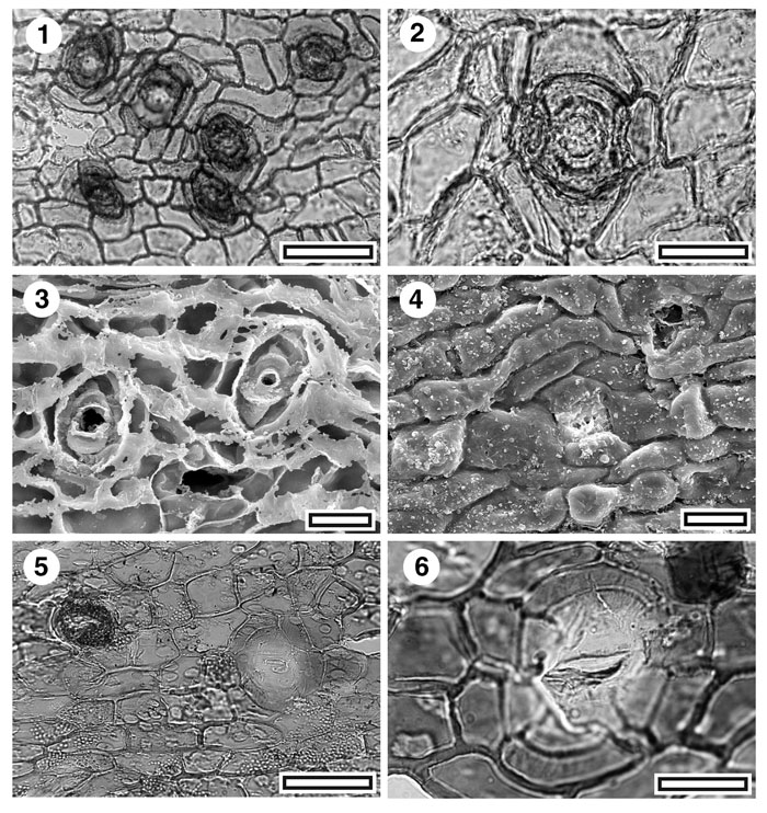
Figure 10. CUT-Mo-EII and CUT-Mo-FEF. 1. CUT-Mo-EII, TLM view of stomatal complexes (SL1873, scale bar = 50 µm); 2. CUT-Mo-EII, TLM detail of stomatal complex (SL1873, scale bar = 20 µm); 3. CUT-Mo-EII, inner SEM detail of two stomatal complexes. Ragged flanges are partial cutinisation of inner periclinal walls (S-1105, scale bar = 20 µm); 4. CUT-Mo-EII, outer SEM view of a single stomatal complex. Note how periclinal walls bulge outwards (S-1105, scale bar = 20 µm); 5. CUT-Mo-FEF, TLM view of stomatal complexes. Note dark ring of trichome at centre left (SL3143, scale bar = 50 µm); 6. CUT-Mo-FEF, TLM detail of stomatal complex (SL3143, scale bar = 20 µm).
|