|
|
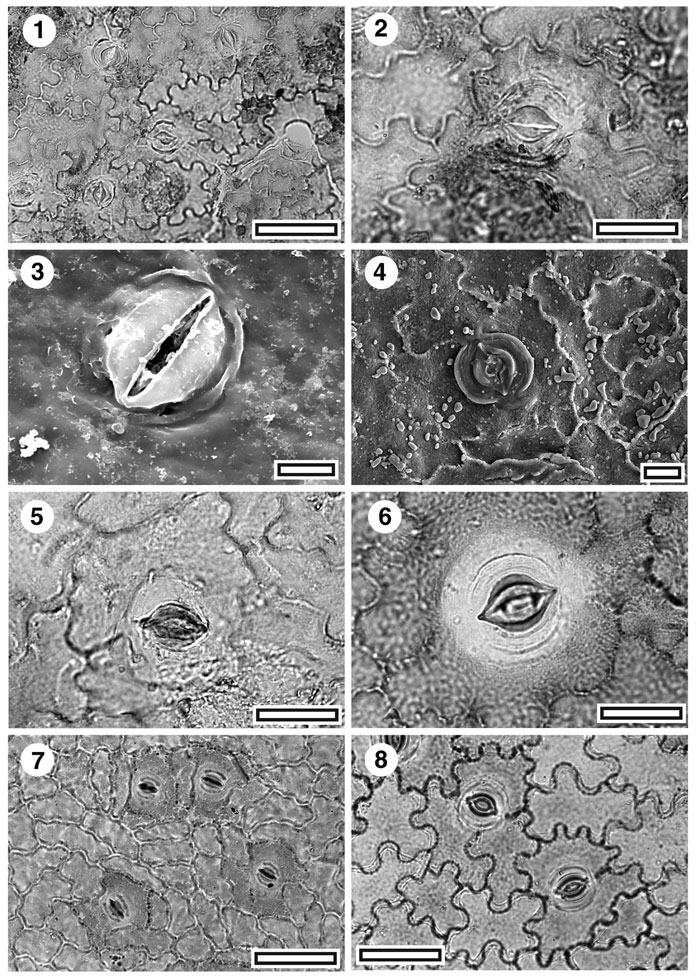
Figure 3. CUT-Mo-EEI, and extant Smilax and Rhipogonum. 1. CUT-Z-EEI, TLM view of stomatal complex (SL5435, scale bar = 50 µm); 2. CUT-Z-EEI, TLM detail of stomatal complex (SL5435, scale bar = 20 µm); 3. CUT-Z-EEI, outer SEM view of a single stoma. Note pinched appearance of outer stomatal ledge, and arching ridges of cuticle (S-1049, scale bar = 10 µm); 4. CUT-Z-EEI, inner SEM view of a single stoma (S-1709, scale bar = 10 µm); 5. CUT-Z-EEI, TLM view of single stomatal complex (SL2035, scale bar = 20 µm); 6. TLM of extant Rhipogonum scandens (OPH2631, scale bar = 20 µm); 7. TLM of extant Smilax australis (AQ037802, scale bar = 50 µm); 8. TLM of extant Rhipogonum album (AQ330750, scale bar = 50 µm).
|