|
|
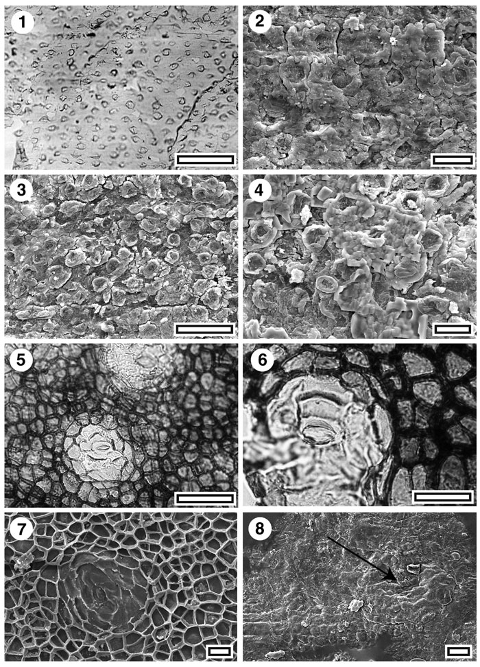
Figure 5. CUT-Mo-EFH and CUT-Mo-FEH. 1. CUT-Mo-EFH, TLM view of rows of papillae (SL1968, scale bar = 50 µm); 2. CUT-Mo-EFH, Outer SEM view of cuticle (S-1059, scale bar = 20 µm); 3. CUT-Mo-EFH, inner SEM view of cuticle (S-1059, scale bar = 50 µm); 4. CUT-Mo-EFH, inner SEM view of cuticle (S-1059, scale bar = 20 µm); 5. CUT-Mo-FEH, TLM view of two stomatal complexes. Note lack of any row structure and complex stomatal construction (SL3238, scale bar = 50 µm); 6. CUT-Mo-FEH, TLM detail of stomatal complex (SL3238, scale bar = 20 µm); 7. CUT-Mo-FEH, Inner SEM view of a single stomatal complex (S-1360, scale bar = 20 µm); 8. CUT-Mo-FEH, Outer SEM view of a single stomatal pore (arrowed) (S-1360, scale bar = 50 µm).
|