|
|
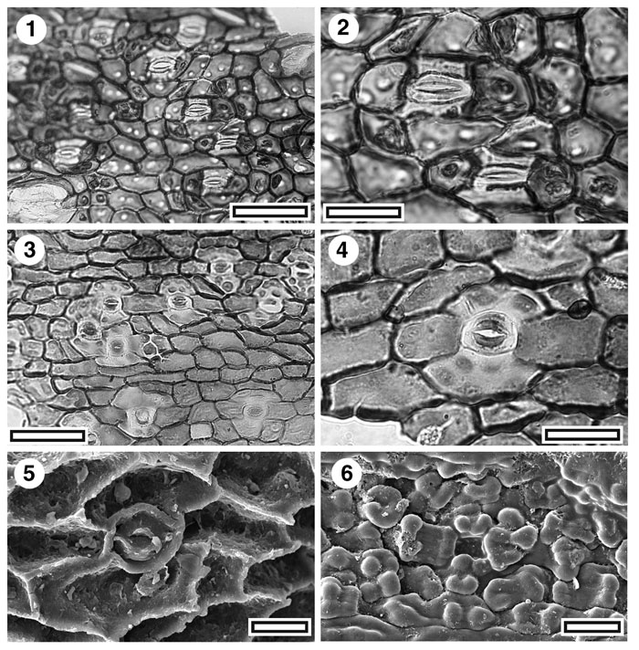
Figure 6. CUT-Mo-FJC and CUT-Mo-FFJ. 1. CUT-Mo-FJC, TLM view of stomatal complexes. Note relatively small and sharply defined papillae (SL2419, scale bar = 50 µm); 2. CUT-Mo-FJC, TLM detail of two stomatal complexes. Note distinctively narrow stoma and relatively small and sharply defined papillae (SL2419, scale bar = 20 µm); 3. CUT-Mo-FFJ, TLM view of stomatal complexes. Note papillae generally confined to subsidiary cells (SL3239, scale bar = 50 µm); 4. CUT-Mo-FFJ, TLM detail of stomatal complex (SL3239, scale bar = 20 µm); 5. CUT-Mo-FFJ, inner SEM detail of a single stomatal complex (S-1361, scale bar = 10 µm); 6. CUT-Mo-FFJ, outer SEM view of at least two stomatal complexes (S-1361, scale bar = 10 µm).
|