|
|
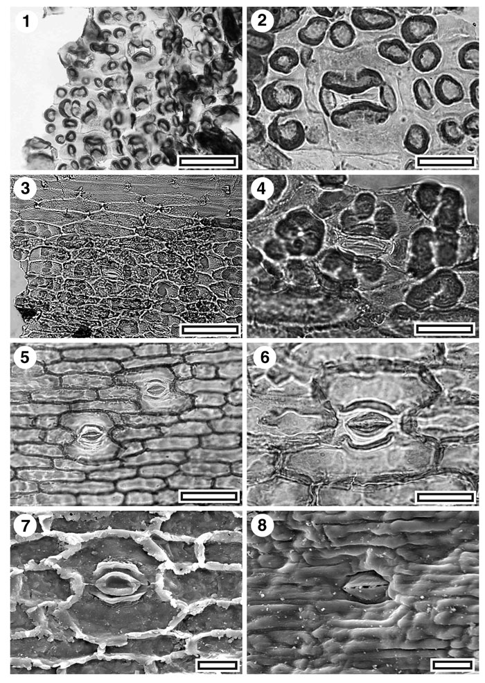
Figure 7. CUT-Mo-FJE, CUT-Mo-AAI, and CUT-Mo-EFI. 1. CUT-Mo-FJE, TLM view of stomatal complexes (SL2524, scale bar = 50 µm); 2. CUT-Mo-FJE, TLM detail of stomatal complex (SL2524, scale bar = 20 µm); 3. CUT-Mo-AAI, TLM view of stomatal complexes, SB1357, scale bar = 50 µm); 4. CUT-Mo-AAI, TLM detail of stomatal complex (SB1357, scale bar = 20 µm); 5. CUT-Mo-EFI, TLM view of two stomatal complexes (SL1973, scale bar = 50 µm); 6. CUT-Mo-EFI, TLM detail of stomatal complex. Note distinctive straight sides to complex and ridges of cuticle flanking the outer stomatal ledges (SL1973, scale bar = 20 µm); 7. CUT-Mo-EFI, inner SEM detail of a single stomatal complex (S-1060, scale bar = 20 µm); 8. CUT-Mo-EFI, outer SEM view of a single stoma. Note pinched appearance of outer stomatal ledge and slit-like aperture (S-1060, scale bar = 20 µm).
|