|
|
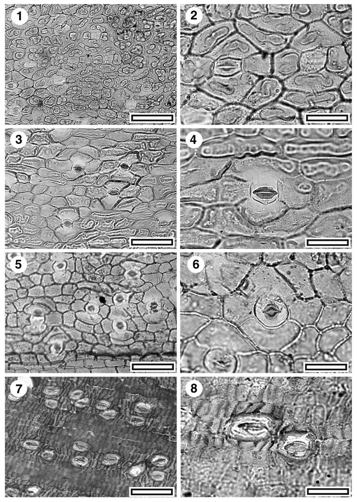
Figure 8. CUT-Mo-EIA, CUT-Mo-FEG, CUT-Mo-EHB, and CUT-Mo-EFF. 1. CUT-Mo-EIA, TLM view of stomatal complexes (SL2446, scale bar = 50 µm); 2. CUT-Mo-EIA, TLM detail of two stomatal complexes (SL2446, scale bar = 20 µm); 3. CUT-Mo-FEG, TLM view of stomatal complexes. Note subdued and fused papillae (SL2093, scale bar = 50 µm); 4. CUT-Mo-FEG, TLM detail of stomatal complex. Note subdued and fused papillae (SL2093, scale bar = 20 µm); 5. CUT-Mo-EHB, TLM view of stomatal complexes. Note absence of any clear row structure or regular costal-intercostal distinction. Some more elongate epidermal cells are visible near the lower edge of the figure (SL1541, scale bar = 50 µm); 6. CUT-Mo-EHB, TLM detail of stomatal complex. Note distinctive rounded outline of the stoma and the pinched ends of the stomatal aperture (SL1541, scale bar = 20 µm); 7. CUT-Mo-EFF, TLM view of stomatal complexes (SL1785, scale bar = 50 µm); 8. CUT-Mo-EFF, TLM detail of two stomatal complexes. Note four isodiametric lateral subsidiary cells on left complex (SL1785, scale bar = 20 µm).
|