|
APPENDIX
Taxonomic Descriptions
 Referred specimens and occurrence: S-283, R-70; SL5381, R-102. Referred specimens and occurrence: S-283, R-70; SL5381, R-102.
Distinguishing features:
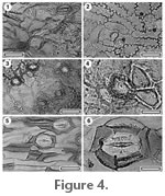
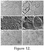
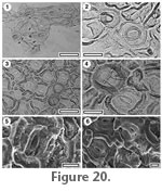
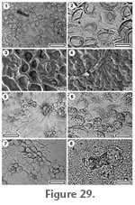
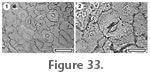
Hill and Pole (1994) described Pterostoma hirsutus from the Cool Store Section. It is distinguished from other Pterostoma by the ornamentation of thick cuticular ridges.
Referred specimens and occurrence: SB0384, R-77; SB0690, R-105.
Distinguishing features: Two specimens have deeply sunken, gymnospermous stomata with some resemblance to Pterostoma, but without the surface ornamentation of P. hirsutus. They are included here for convenience, although they may represent some other form of non-coniferous gymnosperm, like a ginkgophyte.
Referred specimens and occurrence: SL5190, R-04; SL5256, R-13; SL5059, R-21; SL5072, R-23; SL5081, R-24; SL5086, R-25; SL5097, R-26; SL5099, R-27; SL5152, R-32; SL5166, R-33; SL5016, R-61; SL5274, R-68; SL5400, R-74; SB0373, R-75; SL5265, R-76; SL5287; SL5420, R-79; R-104; SL5310, R-105.
Distinguishing features: This cuticle is identified as Bowenia on the basis of having stomates and epidermal cells in rows, stomates with irregularly shaped and typically elongate subsidiary cells, fusiform epidermal cells having periclinal walls of variable thickness (e.g.,
Greguss 1968) and by direct comparison with material published by
Hill (1978), who described Bowenia papillosa and B. eocenica from the Eocene of New South Wales and Victoria. The Tasmanian fossils are identified with B. eocenica on the basis of absence of papillae.
 Reference specimen and occurrence: SL5083, R-25; SL5354, R-30; SL5409, R-34; SL5021, R-68; SL5027, R-71; SL5283, R-104. Reference specimen and occurrence: SL5083, R-25; SL5354, R-30; SL5409, R-34; SL5021, R-68; SL5027, R-71; SL5283, R-104.
Distinguishing features: Agathis is identified on the basis of rounded, coniferous stomatal complexes which are obliquely oriented and under TLM have a strongly thickened ring of cuticle around the stomatal pore, which appears 'suspended' by radiating flanges (Bigwood and Hill 1985;
Hill and Bigwood 1987;
Stockey and Atkinson 1993;
Pole 2007b).
Reference specimen and occurrence: SL5077, R-24; SL5163, R-33.
Distinguishing features: In this study, Araucaria cuticle is recognised as araucarian cuticle with obliquely oriented stomatal complexes and elongate epidermal cells. See
Pole (2000) for further discussion of these features.
Referred specimens and occurrence: SL4995, R-43.
Distinguishing features: A single fragment has typical araucarian structure but with stomatal complexes that are aligned to the long axis of the leaf.
Referred specimens and occurrence: SL5191, R-04; SL5207, R-08; SL5075, R-23; SL5115, R-30 ; SL5178, R-38; SL4997, R-47; SL5279, R-68; SL5026, R-71; SB0331, R-74; SL5033, R-75; SL5263, R-76; SL5290, R-104; SL5308, R-105.
Distinguishing features: In this study, Araucarioides cuticle is recognised as araucarian cuticle with obliquely oriented stomatal complexes and isodiametric epidermal cells. See
Pole (2000) for further discussion.
Bigwood and Hill (1985) described Araucarioides linearis and A. sinuosa from Regatta Point. All material found in this study has straight, rather than sinuous epidermal cell walls. Although sinuous epidermal cells walls were noted in the etymology of A. sinuosa, this featured is not mentioned in the diagnosis and is not apparent in
Bigwood and Hill's (1985) figures.
Reference specimen and occurrence: SL5128, R-30.
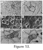
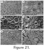
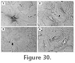
Distinguishing features: Very small scale-like leaves which have monocyclic stomatal complexes and calcium oxalate crystals within the cuticle of the epidermal cells clearly belong in Cupressaceae sensu stricto. They are most likely a species of Libocedrus (Florin and Boutelje 1954). Ruling-out the closely related Austrocedrus (not distinguished from Libocedrus by all botanists) would require more complete material, although there is a broad habitat difference, with Libocedrus predominant in very wet rainforest habitat (Farjon 2005), and Austrocedrus common in drier, sometimes woodland habitats and perhaps with more of a fire-association than Libocedrus (Kitzberger and Veblen. 1999;
Veblen et al. 1999).
Reference specimen and occurrence: SL5122, R-30.
Distinguishing features: This has a very distinctive morphology of monocyclic stomatal complexes, which are in rows and where there is a high degree of networking. Lateral subsidiary cells are often shared between two or even three stomatal complexes in the same row, and subsidiary cells are sometimes shared between stomatal complexes in adjacent rows. These characters identify it as Cupressaceae sensu stricto. A papillate rim around the stomatal aperture is not pronounced and there are no calcium oxalate crystals. However, neither of these characters is ubiquitous within Cupressaceae.
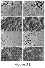
Referred specimens and occurrence: SL5183, R-01; SL5209, R-08; SL5211, R-09; SL5258, R-12; SL5247, R-13; SL5080, R-24; SL5101, R-27; SL5103, R-28; SL5108, R-29; SL5147, R-30; SL5156, R-35; SL5171, R-36; SL5008, R-56; SL5275, R-68; SL5029, R-71; SB0333, R-74; SL5036, R-76.
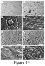
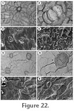
Distinguishing features: Acmopyle has very characteristic stomatal complexes under TLM; the lateral subsidiary cell walls slope gradually from the periclinal to anticlinal position and vary a lot in shape and number (Hill and Carpenter 1991;
Pole 1997a). The polar subsidiary cells are often shared, and there are typically many incompletely formed stomatal complexes. Acmopyle glabra was described from Regatta Point by
Hill and Carpenter
(1991) and has the distinctive stomatal structure of the genus, but lacks the trichomes which are found in some species.
Referred specimens and occurrence: SL5114, R-29; SL5276, R-68.
Distinguishing features: Dacrycarpus has elongated stomatal complexes in chains, which are present in zones of typically only one–three rows, and associated with smooth, elongate epidermal cells (Hill and Carpenter 1991;
Pole 1992).
 Referred specimens and occurrence: SL5110, R-29; SL5131, R-30; SL5414, R-37; SB0356, R-75; SL5261, R-76; SL5303, R-105. Referred specimens and occurrence: SL5110, R-29; SL5131, R-30; SL5414, R-37; SB0356, R-75; SL5261, R-76; SL5303, R-105.
Distinguishing features: Prumnopitys has distinctive stomatal complexes where the subsidiary cells tend to bulge out from their common walls, and a distribution where the nearest neighbouring complex is usually in an adjacent row (Pole 1992,
1997a)
The following taxa are all regarded as most likely Podocarpaceae based on longitudinally-oriented, dicyclic and paratetracyclic stomata (e.g.
Wells and Hill 1989;
Hill and Carpenter 1991;
Hill and Pole 1992;
Hill and Brodribb 1999) and their biogeographic context.
Referred specimens and occurrence: SL5186, R-02; SL5195, R-04; SL5074, R-23; SL5082, R-25; SL5140, R-30; SL5165, R-33; SL5410, R-34; SL5169, R-36; SL5005, R-56; SL5272, R-68; SL5281, R-104; SL5306, R-105.
Distinguishing features: This cuticle type has some resemblance to Acmopyle, but the more crowded bands of stomatal complexes and their more angular outline differ from A. glabra. The generic identification is not certain.
Referred specimens and occurrence: SL5237, R-12.
Distinguishing features: This cuticle morphology has rows of very isodiametric epidermal cells. They may be partially formed stomatal complexes, as found in Acmopyle, but the stomatal form is very distinct.
Referred specimens and occurrence: SL5142, R-30.
Distinguishing features: This cuticle morphology has rows of very circular stomatal complexes in which subsidiary cells are often very thin.
Referred specimens and occurrence: SL5020, R-68.
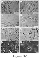
Distinguishing features: The highly sinuous and buttressed epidermal cells of this morphology are very distinctive. The stomatal distribution and, to a lesser degree, outline, suggest Prumnopitys.
Referred specimens and occurrence: SL5214, R-11; SL5240, R-12; SL5091, R-26; SL5351, R-30; SL4994, R-43.
Distinguishing features: This cuticle is recognised by small and narrow stomatal complexes in chains with very broad cells flanking the lateral subsidiary cells, and in zones at least seven stomatal rows wide.
Referred specimens and occurrence: SL5159, R-33; SL5200, R-06; SL5242, R-13; SL5369, R-15; SL5370, R-16.
Distinguishing features: This is a more generalised morphological of Podocarpaceae cuticle with more isodiametric epidermal cells and with a distinctively thickened ring of cuticle around the stomatal pore. Further study may show that some of the specimens listed above come from distinct taxa.
Referred specimens and occurrence: SL5132, R-30.
Distinguishing features: The distinctive feature of this cuticle is the papillae found on the subsidiary and epidermal cells. In this respect and in general morphology it resembles Kahahuia (Pole 1997b), which has been placed in the Taxaceae (Pole 2007b). Similar material was illustrated by
Carpenter et al. (2004).
 Reference specimen and locality: SL5298, R-104. Reference specimen and locality: SL5298, R-104.
Referred specimens and occurrence: SL5187, R-02; SL5189, R-03; SL5205, R-07; SL5111, R-29; SL5145, R-30; SL5153, R-32; SL4993, R-42; SL5018, R-68.
Stomatal complexes. Stomatal distribution over leaf surfaces unknown, stomata evenly spread on stomatal surface, isolated, randomly oriented, essentially brachyparacytic, but also with subsidiary cells modified by a tangential division, giving two lateral and two polar cells at right angles to stomatal axis, size range unimodal, at same level as normal epidermal cells. Subsidiary cell periclinal walls same thickness as normal epidermal cells. Guard cell pair outline distinctly elongate-rectangular, outlined by a well-defined anticlinal wall, length 27–37 µm (medium), at same level as subsidiary cells (exposed on surface), little polar development between guard cells (guard cells appear as continuous ring). Outer stomatal ledge not apparent, outermost cuticle possibly lying directly over guard cells, much thinner than normal epidermal cells, often broken away, with a slit-like pore.
Epidermal cells. Epidermal cell flanges clearly visible using TLM, normal cells typically elongate, larger than the stomata, cells over veins not differentiated, anticlinal walls wavy, unbuttressed, unornamented.
Indumentum. Glabrous.
Distinguishing features. Distinguished by the distinctive shape of the guard cell pair; elongated rectangles, with no division at the polar ends, with very thin cuticle overlying them, and a slit-like pore.
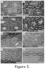
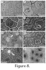
Identification. This very distinctive shape of the guard cells links this taxon with Gnetum in the Gnetaceae (Figures 7.2, 7.4, and see
Paliwal et al. 1974;
Nautiyal et al. 1976). However, all the extant Gnetum appear to have purely brachyparacytic stomatal complexes, whereas the fossil commonly has a pair of polar subsidiary cells as well as a pair of lateral subsidiary cells. This suggests CUT-Z-GDB represent an extinct genus of Gnetaceae.
 Reference specimen and locality: SB0756, R-102. Reference specimen and locality: SB0756, R-102.
Referred specimens and occurrence: SB0360, R-75.
Stomatal complexes. Stomatal distribution over leaf surfaces unknown, stomata evenly spread, isolated, randomly oriented, brachyparacytic, size range unimodal. Subsidiary cells typically elongate tangential to stomata and often longer than the stomata, periclinal walls of same thickness as normal epidermal cells, unornamented. Guard cell pair outline elliptical, outlined by a well-defined anticlinal wall, length 25-35 µm (medium). Outer stomatal ledge elliptical, extending from outer edge of stoma, same thickness as normal epidermal cells, pore elliptical.
Epidermal cells. Epidermal cell flanges clearly visible using TLM, highly variable from isodiametric to elongate, approximately the same size, or slightly smaller than the stomata, anticlinal walls straight to curved, unbuttressed, coarsely granular, unornamented.
Indumentum. Glabrous. Lid-cells present, consisting of four cells slightly smaller than normal epidermal cells, but with thinner cuticle.
Distinguishing features. Paracytic subsidiary cells and coarsely granular cuticle.
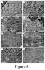
Identification. The brachyparacytic stomatal complexes, prominent outer stomatal ledges and coarsely granular epidermal cells suggest this as Winteraceae (Figures 8.6-8.8), although the lack of granular plugging of the stomatal pore (typical of extant species) introduces an element of doubt. One of the only two specimens (SB0360) has what is interpreted as a four-parted complex of lid cells (forming a lid over a gland). There is no ridge or scar on this structure which might indicate it was some form of trichome base. The presence of lid cells is intriguing, only one taxon of Winteraceae is known to me to have lid cells, Bubbia semecarpoides, but these are single-celled.
Parallel-veined monocot cuticle is identified on the basis of stomatal complexes which are aligned along the long axis of the leaf, and typically have a paratetracytic structure, or some derivative of it (Stebbins and Khush 1961; Tomlinson 1974; Dahlgren and Clifford 1982). Some conifers have this structure but differ from monocots in having guard cells overarched by the subsidiary cells. The cuticle of the net-veined monocots is fundamentally different and is identified by direct comparison with living taxa.
Key to Parallel-veined Monocot cuticle
1. Trichome bases present. CUT-Z- JJG (Arecaceae)
1. Trichome bases absent. 2.
2. Cuticle very thin, outer stomatal ledges thickest part of cuticle. CUT-Z-JBC
2. Stomatal complexes large and subsidiary cell cuticle robustly thickened. CUT-Mo-GCE
 Reference specimen and locality: SB0388, R-75. Reference specimen and locality: SB0388, R-75.
Stomatal complexes. Stomatal distribution over leaf surfaces unknown, stomata evenly spread, isolated, randomly oriented, brachyparacytic, size range unimodal. Subsidiary cells irregularly shaped, periclinal walls same thickness as normal epidermal cells, ornamented with a more or less continuous ridge flanking the outer stomatal ledge. Guard cell pair outline circular, outer margin obscured under TLM by surface ornamentation, length 25–33 µm (medium), little polar development between guard cells (guard cells appear as continuous ring). Outer stomatal ledge a distinctive 'lemon' shape, broad in the middle, and narrowing sharply at either end, thicker than normal epidermal cells, extending over inner edge of stoma, pore elliptical - subcircular.
Epidermal cells. Epidermal cell flanges clearly visible using TLM, normal cells highly variable from isodiametric to elongate, typically slightly larger than the stomata, cells over major veins more elongate, anticlinal walls markedly sinuous, unbuttressed, unornamented.
Indumentum. Glabrous.
Distinguishing features. Brachyparacytic stomatal complexes with highly sinuous epidermal cell walls.
Identification: The cuticle is identified as Rhipogonum based firstly on the distinctive brachyparacytic stomatal complexes, and highly sinuous epidermal cell walls. The rather similar Smilax tends to have smaller stomata and more diffuse epidermal cell walls, although there is some overlap. A very similar form of cuticle is present on Early Miocene leaves in New Zealand with Rhipogonum leaf architecture (Pole 1996). Along with impressions of the family from Melville Island, furthest north Australia, which are likely of early Tertiary age (Pole 1998c) these are the earliest record of the family in Australia.
Reference specimen and locality: SB0744, R-70.
Stomatal complexes. Stomatal distribution over leaf surfaces unknown, isolated, aligned parallel to long axis of leaf, brachyparacytic, size range unimodal. Subsidiary cells typically elongate tangential to stomata, periclinal walls same thickness as normal epidermal cells, ornamented by a ridge above the distal wall. Guard cell pair outline narrowly elliptic, outlined by a well-defined anticlinal wall, length 20–23 µm (medium), little polar development between guard cells (guard cells appear as continuous ring). Outer stomatal ledge not clear, same thickness as normal epidermal cells, pore elliptical.
Epidermal Cells. Epidermal cell flanges somewhat diffuse, approximately the same size, or slightly smaller than the stomata, anticlinal walls sinuous, unbuttressed, unornamented.
Indumentum. With sparse scars of trichome bases (trichomes deciduous), inserted between epidermal cells, with a thickened poral rim.
Distinguishing features. Monocot cuticle with trichome insertion scars with smooth, thickened poral rims.
Identification. Based on the trichome insertion scars this is regarded as Arecaceae.
Reference specimen and locality: SB0393, R-75.
Stomatal complexes. Stomatal distribution over leaf surfaces unknown, stomata evenly spread, isolated, showing a clear trend towards alignment, size range unimodal. Subsidiary cells difficult to count under TLM, periclinal walls same thickness as normal epidermal cells. Guard cell pair outline difficult to distinguish, length 25–28 µm (medium). Outer stomatal ledge narrowly elliptic, thicker than normal epidermal cells, extending over inner edge of stoma, pore slit-like.
Epidermal Cells. Epidermal cell flanges very thin or absent (anticlinal walls of epidermal cells not clear under TLM), normal cells elongated, anticlinal walls straight to curved, unbuttressed, unornamented.
Indumentum. Glabrous.
Distinguishing features. Monocot cuticle which is very thin with only the narrowly elliptic outer stomatal ledges being prominent. Sometimes with single ridges of cuticle flanking the outer stomatal ledges.
 Reference specimen: SL4999, R-47. Reference specimen: SL4999, R-47.
Referred specimens and occurrence: SL5199, R-06; SL5238, R-12; SL5255, R-13; SL5052, R-21; SL4999, R-47; SL5430, R-74; SL5425, R-79.
Description. Epidermis not divided into costal and intercostal zones (stomata evenly spread). Stomatal complexes paratetracytic, with four subsidiary cells (two polar and two lateral, the lateral subsidiary cells, or both the lateral and polar subsidiary cells have much thicker anticlinal walls than normal epidermal cells), with a typically angular outline, not in rows or chains, but widely scattered (nearest neighbour often to the side), longitudinally oriented. Subsidiary cells with periclinal walls thinner than those of normal epidermal cells, not papillate. Outer stomatal ledge 30–43 µm long, distinctly elliptical, with elongate aperture, cuticle thicker than normal epidermal cells. Epidermal cells clearly visible under TLM, in clear rows, elongate, straight-walled, unbuttressed, glabrous, not papillate.
Distinguishing features. Typical monocot cuticle but with subsidiary cells with prominently thickened anticlinal walls.
Lauraceae cuticle is identified on the basis of paracytic stomatal complexes with guard cells embedded within the subsidiary cells, and the presence of cuticular scales, or flanges between them (Bandulska 1926,
Hill 1986,
Christophel et al. 1996,
Vadala and Greenwood 2001). Lauraceae have simple, deciduous, trichomes with poral bases. This clearly distinguishes them from Myristicaceae which have a similar stomatal structure, but multi-celled trichome bases (Upchurch and Dilcher 1990, Pole, pers. obs.) Identification with extant genera of Australasian Lauraceae is based on
Christophel and Rowett (1996).
1. Stomatal complexes aligned. CUT-L-DDE
1. Stomatal complexes randomly oriented. 2.
2. Surface ornamented with prominent striations. CUT-L-DDC
2. No striations. 3.
3. Papillae present. 4.
3. Papillae not present. 5.
4. Whole surface of epidermal cells surfaces raised in "bubble-like" fashion. CUT-L-JEC
4. Papillae distinct, with the margins of the epidermal cell. CUT-L-GCI
5. Anticlinal walls of subsidiary cells and those cells in contact with subsidiaries are markedly thickened. CUT-L-DCG
5. Anticlinal wall thickness of subsidiary and ordinary epidermal cells similar. 6.
6. Epidermal cells sinuous or markedly wavy. CUT-L-DCD
6. Epidermal cells polygonal or only slightly wavy. 7.
7. Epidermal cells immediately surrounding stomatal complex typical in an anisocytic pattern (and often stain slightly darkly than normal epidermal cells). CUT-L-DCI
7. Epidermal cells immediately surrounding stomatal complex not in any discernable pattern (and all epidermal cells staining similarly). 8.
8. Subsidiary cells of distinctly different thickness (staining differently) than normal epidermal cells. 9.
8. Subsidiary cells staining similarly to or less than epidermal cells. 10.
9. Subsidiary cells staining less than epidermal cells. 14.
9. Subsidiary cells staining more than epidermal cells, trichome bases present and strongly thickened. CUT-L-DCF
10. Cuticular scales distinctly 'double'. CUT-L-DDJ
10. Cuticular scales not 'double'. 11.
11. Stomatal complex large. CUT-L-DCH
11. Stomatal complex small-medium. 12.
12. Stomatal complex medium, cuticle of moderate thickness.
12. Stomatal complexes small, cuticle very thin. 13.
13. Outline of stomatal complex slightly overgrown by cuticle. CUT-L-JBI
13. Outline of stomatal complex not overgrown by cuticle. CUT-L-JBA
14. Essentially glabrous. CUT-L-GCG
14. With prominent trichome insertion scars. CUT-L-GCH
 Reference specimen and locality: SB0730, R-16. Reference specimen and locality: SB0730, R-16.
Stomatal complexes. Stomatal distribution over leaf surfaces unknown, evenly spread, isolated, transversely oriented, paracytic, outline typically broader than long, length 28–38 µm (medium). Subsidiary cell periclinal cuticle thinner than over normal epidermal cells. Cuticular scales narrow.
Epidermal Cells. Epidermal cell flanges clearly visible using TLM, cells over veins not distinguished by shape, normal epidermal cells isodiametric, walls straight, unbuttressed.
Indumentum. Glabrous, unornamented.
Distinguishing features. Lauraceae cuticle with stomatal complexes transverse to the long axis of the leaf.
Reference specimen and locality: SB0670, R-23.
Stomatal complexes. Stomatal distribution over leaf surfaces unknown, Stomatal complexes evenly spread, isolated, randomly oriented, paracytic, outline polygonal, length 28–38 µm (medium). Subsidiary cell periclinal cuticle thinner than over normal epidermal cells. Cuticular scales narrow.
Epidermal Cells. Epidermal cell flanges clearly visible using TLM, cells over fine venation poorly distinguished as 'venal' (elongated), normal epidermal cells isodiametric, walls straight to curved, unbuttressed.
Indumentum. With scars of trichome bases, ornamented with bands of ridges joining some stomatal complexes and radiating from trichome bases. Trichome insertion scars common (trichomes deciduous and therefore trichome type unknown), inserted between epidermal cells, modified with thickened poral rim and radial walls. Epidermal cells around trichome scar form a distinct ring of isodiametric foot cells.
Distinguishing features. Lauraceae cuticle with an ornamentation of ridges and with very thickened trichome insertion poral rims.
 Reference specimen and locality: SL0719, R-102. Reference specimen and locality: SL0719, R-102.
Stomatal complexes. Stomatal distribution over leaf surfaces unknown, Stomatal complexes in clear areoles, isolated, randomly oriented, paracytic. Cuticular scales not clear.
Epidermal Cells. Epidermal cell flanges generally hidden by papillae, cells over fine venation distinguished as 'venal' (elongated), normal epidermal cells isodiametric, walls curved or wavy, unbuttressed.
Indumentum. With trichome insertion scars and papillose, unornamented. Papillae present over all epidermal cells, formed by the entire outer surface of the epidermal cell projecting upwards in a bubble-like fashion, smooth. Trichome insertion scars common (trichomes deciduous and therefore trichome type unknown), inserted between epidermal cells. Epidermal cells around trichome scar radially elongated as distinct foot cells, unthickened.
Distinguishing features. Papillate Lauraceae cuticle with bubble-like papillae and with trichome scars.
Reference specimen and locality: SL5372, R-102.
Stomatal complexes. Stomatal distribution over leaf surfaces unknown, Stomatal complexes in clear areoles, isolated, randomly oriented, paracytic length 15-18 µm (medium). Cuticular scales not clear.
Epidermal cells. Epidermal cell flanges not in TLM (cuticle thin), cells over fine venation distinguished as 'venal' (elongated), normal epidermal cells isodiametric, walls curved or wavy, unbuttressed.
Indumentum. With trichome insertion scars and papillose, unornamented. Papillae present over all epidermal cells, smooth, discrete, about one half to two thirds of the diameter of the epidermal cell. Trichome insertion scars common (trichomes deciduous and therefore trichome type unknown), inserted between epidermal cells. Epidermal cells around trichome scars radially elongated as distinct foot cells, with thickened radial walls and pore.
Distinguishing features. Papillate Lauraceae cuticle with discrete papillae.
 Reference specimen and locality: SB0346, R-74. Reference specimen and locality: SB0346, R-74.
Referred specimens and occurrence: SL5198, R-06; SL5215, R-11; SL5223, R-12; SL5049, R-21; SL5105, R-28; SL5017, R-61; SB0346, R-74; SB0352, R-75; SL5421, R-79; SL5270, R-103.
Stomatal complexes. Stomatal distribution over leaf surfaces unknown. Stomatal complexes evenly spread, isolated, randomly oriented, paracytic, length 18–23 µm (medium). Subsidiary cell anticlinal walls, including walls forming the stomatal pore, markedly thickened, periclinal cuticle not distinct in thickness from normal epidermal cells. Cuticular scales narrow.
Epidermal cells. Epidermal cell flanges clearly visible using TLM, cells over veins not distinguished by shape, normal epidermal cells isodiametric, walls straight to curved, unbuttressed. Epidermal cells near the stomatal complex may have thickened anticlinal walls.
Indumentum. Persistent uniseriate trichomes inserted over 1-3 epidermal cells.
Distinguishing features. Lauraceae cuticle readily distinguished by the remarkable thickening of anticlinal walls within and sometimes around the stomatal complex.
 Reference specimen and locality: SB0349, R-74. Reference specimen and locality: SB0349, R-74.
Referred specimens and occurrence: SL5098, R-27.
Stomatal complexes. Stomatal distribution over leaf surfaces unknown, Stomatal complexes in clear areoles, isolated, randomly oriented, paracytic, outline rounded, but irregular, length 13–30 µm, (small-medium). Subsidiary cell periclinal cuticle not distinct in thickness from normal epidermal cells. Cuticular scales 'butterfly-like'.
Epidermal cells. Epidermal cell flanges clearly visible using TLM, cells over fine venation distinguished as 'venal' (elongated), normal epidermal cells highly variable from isodiametric to elongate, walls sinuous, unbuttressed.
Indumentum. With trichome insertion scars, unornamented. Trichome scars sparse (trichomes deciduous), inserted between epidermal cells, Epidermal cells around trichome scar radially elongated as distinct foot cells, unthickened.
Distinguishing features. Lauraceae cuticle with sinuous epidermal cell anticlinal walls and butterfly-like scales.
Reference specimen and locality: SB0672, R-102.
Referred specimens and occurrence: SL5332, R-20; SL5343, R-23; SL5084, R-25; SB0672, R-27; SB0399, R-75; SL5432, R-102; SL5313, R-105.
Stomatal complexes. Stomatal distribution over leaf surfaces unknown, Stomatal complexes evenly spread, isolated, randomly oriented, paracytic, outline rounded but with flattened poles, length 15–25 µm (medium). Subsidiary cell periclinal cuticle thicker than over normal epidermal cells. Cuticular scales narrow.
Indumentum. With trichome insertion scars, unornamented. Trichome scars common, (trichomes deciduous), inserted between epidermal cells. Epidermal cells around trichome scar modified to form a thickened poral rim.
Epidermal cells. Epidermal cell flanges clearly visible using TLM, cells over fine venation distinguished as 'venal' (elongated), normal epidermal cells highly variable from isodiametric to elongate, walls straight to curved, unbuttressed. Cells immediately around the paracytic stomatal, complex are arranged in an anisocytic manner, with three and sometimes four cells.
Distinguishing features. Lauraceae cuticle with cells around the stomatal complex arranged in an anisocytic pattern.
 Reference specimen and locality: SB0666, R-22. Reference specimen and locality: SB0666, R-22.
Referred specimens and occurrence: SL5045, R-21; SB0731, R-22; SL5341, R-23; SB0348, R-74; SL5271, R-103.
Stomatal complexes. Stomatal distribution over leaf surfaces unknown, Stomatal complexes evenly spread, isolated, randomly oriented, paracytic, outline rounded but with flattened poles, length 15–23 µm (medium). Subsidiary cell periclinal cuticle markedly thicker than over normal epidermal cells. Cuticular scales double.
Epidermal cells. Epidermal cell flanges clearly visible using TLM, cells over fine venation distinguished as 'venal' (elongated), normal epidermal cells highly variable from isodiametric to elongate, walls straight to curved, unbuttressed.
Indumentum. With trichome insertion scars, unornamented. Trichome scars common, (trichomes deciduous), inserted between epidermal cells. Epidermal cells around trichome scar modified to form thickened poral rim and radial walls.
Distinguishing features. Lauraceae cuticle with periclinal walls of subsidiary cells distinctly thicker than normal epidermal cells, and with massively thickened poral rims of trichome insertion scars.
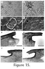
Identification: Although the stomatal structure is entirely consistent with, and in terms of extant plants, unique to Lauraceae, several leaf fragments indicate that the margin was toothed (Figure 15.5-15.8). Teeth are unknown in extant Lauraceae (Sassafras is lobed). It is possible that Lauraceae once included taxa with teeth, and it is also possible that this represents a related, but extinct family. For convenience, the cuticle is included here as Lauraceae. The 'double' cuticular scales recall Endiandra (Christophel and Rowett 1996) but the shape of the stomatal complexes and the thicker subsidiary cell cuticle are further evidence of at least generic difference.
 Reference specimen and locality: SB0398, R-75. Reference specimen and locality: SB0398, R-75.
Referred specimens and occurrence: SL5319, R-06; SL5331, R-20; SL5094, R-26; SB0398, R-75; SL5305, R-105.
Stomatal complexes. Stomatal distribution over leaf surfaces hypostomatic, Stomatal complexes evenly spread, isolated, randomly oriented, paracytic, outline rounded but with flattened poles, length 18–23 µm (medium). Subsidiary cell periclinal cuticle thinner than over normal epidermal cells. Cuticular scales double.
Epidermal cells. Epidermal cell flanges clearly visible using TLM, cells over fine venation distinguished as 'venal' (elongated), normal epidermal cells highly variable from isodiametric to elongate, walls curved or wavy, unbuttressed.
Indumentum. With trichome insertion scars, unornamented. Trichome scars common, (trichomes deciduous), inserted between epidermal cells. Epidermal cells around trichome scar irregularly elongated as foot cells, unthickened.
Non-stomatal surface. Non-Stomatal Surface Epidermal cells isodiametric, wavy and slightly buttressed. Simple trichome insertion scars present.
Distinguishing features. Lauraceae cuticle with 'double' cuticular scales and slightly buttressed epidermal cells.
Identification. The 'double' cuticular scales, stomatal complex outline and thinner subsidiary cell cuticle than epidermal cells is completely consistent with this being an Endiandra (Christophel and Rowett 1996).
Reference specimen and locality: SB0728, R-6.
Stomatal complexes. Stomatal distribution over leaf surfaces unknown, Stomatal complexes evenly spread, isolated, randomly oriented, paracytic, outline rounded but with flattened poles, length c. 45 µm (medium). Subsidiary cell periclinal cuticle not distinct in thickness from normal epidermal cells. Cuticular scales 'butterfly-like'.
Epidermal cells. Epidermal cell flanges clearly visible using TLM, cells over veins not distinguished by shape, normal epidermal cells isodiametric, walls straight, unbuttressed.
Indumentum. Glabrous, unornamented.
Distinguishing features. Lauraceae cuticle with large stomatal complexes.
Note. The apparent cuticular scales on this taxon may be an artefact of preservation. The stomatal complex size is unusually large for Lauraceae. It is placed here until further and perhaps better preserved specimens clarify this point.
 Reference specimen and locality: SB0700, R-102. Reference specimen and locality: SB0700, R-102.
Referred specimens and occurrence: SL5028, R71.
Stomatal complexes. Stomatal distribution over leaf surfaces unknown, Stomatal complexes in clear areoles, isolated, randomly oriented, paracytic, outline rounded, but irregular, length 13–18 µm, (small-medium). Subsidiary cell periclinal cuticle not distinct in thickness from normal epidermal cells, anticlinal walls particularly strong and in TLM view appear to slightly overhang the stomatal complex. Cuticular scales butterfly-like.
Epidermal cells. Epidermal cell flanges not clearly visible under TLM, cells over fine venation distinguished as 'venal' (elongated), normal epidermal cells highly variable from isodiametric to elongate, walls curved or wavy, unbuttressed.
Indumentum. With trichome insertion scars, unornamented. Trichome scars common, (trichomes deciduous), inserted between epidermal cells. Epidermal cells around trichome base radially elongated to form distinct foot cells and thickened to form a poral rim.
Distinguishing features. Lauraceae cuticle which is very thin, and with stomatal complexes sunken into individual pits and surrounded by an irregular rim.
Reference specimen and locality: SB0391, R-75.
Referred specimens and occurrence: SL5065, R-21; SL5345, R-23.
Stomatal complexes. Stomatal distribution over leaf surfaces unknown, Stomatal complexes evenly spread, isolated, randomly oriented, paracytic, outline rounded, but irregular, length 25–28 µm (medium). Subsidiary cell periclinal cuticle not distinct in thickness from normal epidermal cells. Cuticular scales narrow.
Epidermal cells. Epidermal cell flanges very thin or absent (anticlinal walls of epidermal cells not clear under TLM), cells over fine venation distinguished as 'venal' (elongated), normal epidermal cells unclear, walls curved or wavy, unbuttressed.
Indumentum. Glabrous, unornamented.
Distinguishing features. Very thin Lauraceae cuticle.
 Reference specimen and locality: SL1023, R-21. Reference specimen and locality: SL1023, R-21.
Referred specimens and occurrence: SL5324, R-06; SL5228, R-12; SL5244, R-13; SL5269; SL5423, R-79; R-103; SL5307, R-105.
Stomatal complexes. Stomatal distribution hypostomatic, stomatal complexes evenly spread, isolated, randomly oriented, paracytic, outline rounded but with flattened poles, length 15–25 µm (medium). Subsidiary cell periclinal cuticle thinner than over normal epidermal cells (occasionally not apparent). Cuticular scales narrow, but with prominent triangular flaps of cuticle at the polar ends.
Epidermal cells. Epidermal cell flanges clearly visible using TLM, cells over fine venation distinguished as 'venal' (elongated), normal epidermal cells highly variable from isodiametric to elongate, walls straight to curved, unbuttressed.
Indumentum. Essentially glabrous, but very rarely simple, poral trichome insertion scars present.
Distinguishing features. Lauraceae cuticle with subsidiary cell cuticle thinner than normal epidermal cell cuticle and glabrous. The basic form and size of the stomatal complexes is rather similar to CUT-L-DCI, but lacks the anisocytic pattern of epidermal cells around the stomatal complex.
Reference specimen and locality: SL5374, R-102.
Stomatal complexes. Stomatal distribution unknown, stomatal complexes in distinct, small clusters within areoles, isolated, randomly oriented, paracytic, outline rounded but with flattened poles, length 15–25 µm (medium). Subsidiary cell periclinal cuticle much thinner than over normal epidermal cells. Cuticular scales butterfly-like.
Epidermal cells. Epidermal cell flanges clearly visible using TLM, cells over fine venation distinguished as 'venal' (slightly elongated), normal epidermal cells highly variable from isodiametric to elongate, walls straight to curved, unbuttressed.
Indumentum. With trichome insertion scars, unornamented. Trichome scars common, (trichomes deciduous), often inserted over the junction of three epidermal cells. Epidermal cells around trichome base radially elongated to form distinct foot cells and slightly thickened to form a poral rim and radiating walls.
Distinguishing features. Lauraceae cuticle with subsidiary cell cuticle thinner than normal epidermal cell cuticle and with prominent trichome insertion scars.
Proteaceae cuticle is identified on the basis of brachyparacytic stomatal structure and rounded trichome scars, at leats some of which overlie more than one epidermal cell (Lange 1978,
Carpenter 1994,
Carpenter et al. 2004).
Key to Proteaceae cuticle
1. Cuticle with prominent surface striations. 2.
1. Cuticle without striations, or striations subdued. 4.
2. Epidermal cell walls sinuous. CUT-P-EJG
2. Epidermal walls curved or indistinct. 3.
3. Trichome base outline diffuse, surface ornamentation of striations in flowing bands. CUT-P-EJF
3. Trichome base outline well-defined, surface ornamentation of ridges above epidermal cell anticlinal walls. CUT-P-EJE
4. Epidermal surface ornamented with papillae. 5.
4. Epidermal surface smooth or without prominent ridging. 6.
5. Papillae elongate. CUT-P-EJH
5. Papillae circular and with a diameter distinctly smaller than their epidermal cell. CUT-P-GDJ
6. Trichome bases prominent, stomata with random orientation. 7.
6. Trichome bases not prominent, stomata with a trend towards alignment. CUT-P-EAA
6. Trichome bases surrounded by radially elongate cells, epidermal cell walls clear, Prominent T-pieces at guard cell poles. Stomatal complexes generally parallel CUT-P-EJI
6. Trichome bases not surrounded by radially elongate cells, epidermal cell walls often indistinct, No T-pieces, epidermal cells polygonal. CUT-P-EJD
 Reference specimen and locality: SB0674, R-104. Reference specimen and locality: SB0674, R-104.
Referred specimens and occurrence: SL5361, R-20; SL5050, R-21; SB0750, R-23; SL5007, R-56; SL5277, R-68; SB0389, R-75; SL5264, R-76.
Stomatal complexes. Stomatal distribution over leaf surfaces unknown, stomata evenly spread, isolated, randomly oriented, brachyparacytic, size range unimodal. Subsidiary cells typically elongate tangential to stomata, periclinal walls same thickness as normal epidermal cells, ornamented with many fine ridges parallel with, and on either side of the stomata. Guard cell pair outline elliptical, length 25–33 µm (medium), with prominent polar rods. Outer stomatal ledge elliptical, extending from outer edge of stoma, thicker than normal epidermal cells, pore elliptical.
Epidermal cells. Epidermal cell flanges clearly visible using TLM, normal cells elongated, approximately the same size as the stomata, cells over veins not distinguished by shape, anticlinal walls markedly sinuous; slightly buttressed, ornamented with 'flowing' pattern of many prominent ridges.
Indumentum. With trichome insertion scars, and basal part of trichome persistent. Trichome scars common, annular and multicellular, inserted over 1–2 modified epidermal cells, diameter similar in size to normal epidermal cell. Epidermal cells under trichome base modified to form a thick, raised circular platform, on top of which sits a smooth, thick hollow collar.
Distinguishing features. Proteaceae cuticle with prominent surface striations and persistent trichomes.
Reference specimen and locality: SB0679, R-6.
Referred specimens and occurrence: SB0740, R-25.
Stomatal complexes. Stomatal distribution over leaf surfaces unknown, stomata evenly spread, isolated, randomly oriented, brachyparacytic, size range unimodal. Subsidiary cell number and shape unclear. Guard cell pair outline elliptical, not outlined by a clear anticlinal wall, length about 20 µm (medium), little polar development between guard cells (guard cells appear as continuous ring). Outer stomatal ledge elliptical, extending from outer edge of stoma, same thickness as normal epidermal cells, pore elliptical.
Epidermal cells. Epidermal cell flanges from clearly defined to indistinct, normal cells highly variable from isodiametric to elongate, approximately the same size as the stomata, cells over veins not distinguished by shape, anticlinal walls curved, unbuttressed, ornamented with 'flowing' pattern of many prominent ridges.
Indumentum. With trichome insertion scars (trichomes deciduous). Trichome scars sparse, annular and multicellular, inserted over 1–2 modified epidermal cells, diameter similar in size to normal epidermal cell. Epidermal cells under trichome base modified to form a thick, raised circular platform, on top of which sits a smooth, thick hollow collar.
Distinguishing features. Proteaceae cuticle which is thin, but with prominently thickened trichome bases and an ornamentation of flowing ridges.
 Reference specimen and locality: SB0680, R-70. Reference specimen and locality: SB0680, R-70.
Referred specimens and occurrence: SL5068, R-21; SL5006, R-56; SB0680, R-70.
Stomatal complexes. Stomatal distribution over leaf surfaces unknown, stomata evenly spread, isolated, showing a clear trend towards alignment, brachyparacytic, size range unimodal. Subsidiary cells typically elongate tangential to stomata, periclinal walls same thickness as normal epidermal cells, ornamented with 1–2 ridges parallel with, and on either side of the stomata. Guard cell pair outline elliptical, outlined by a well-defined anticlinal wall, length 23–25 µm (medium), clearly separated by polar walls. Outer stomatal ledge elliptical, extending from outer edge of stoma, same thickness as normal epidermal cells, pore elliptical - sub circular.
Epidermal cells. Epidermal cell flanges distinct (although may be partly obscured by surface topography), normal cells elongated, approximately the same size as the stomata, cells over veins not distinguished by shape, anticlinal walls curved, unbuttressed, ornamented with strong ridges above the epidermal cell anticlinal walls.
Indumentum. With trichome insertion scars (trichomes deciduous). Trichome bases common, annular and multicellular, inserted over 1–2 modified epidermal cells. Diameter similar in size to normal epidermal cell. Epidermal cells under trichome base modified to form a thick, raised circular platform, on top of which sits a smooth, thick hollow collar. Trichome platform is granular and frilled with a markedly thickened collar.
Distinguishing features. Proteaceae cuticle with pronounced ridges over the epidermal cell anticlinal walls.
Reference specimen and locality: SB0677, R-76.
Stomatal complexes. Stomatal distribution over leaf surfaces unknown, stomata evenly spread, isolated, randomly oriented, brachyparacytic, size range unimodal. Subsidiary cells typically elongate tangential to stomata, periclinal walls same thickness as normal epidermal cells, unornamented. Guard cell pair outline elliptical, outlined by a well-defined anticlinal wall, length 20–25 µm (medium), little polar development between guard cells (guard cells appear as continuous ring). Outer stomatal ledge elliptical, extending from outer edge of stoma, thinner than normal epidermal cells, pore elliptical.
Epidermal cells. Epidermal cell flanges distinct (although may be partly obscured by surface topography), normal cells elongated, typically as long as, although narrower than typical epidermal cells, cells over veins not distinguished by shape, anticlinal walls curved, unbuttressed, unornamented.
Indumentum. With trichome insertion scars (trichomes deciduous) and papillae. Trichome scars common, annular and multicellular, inserted over 2–3 modified epidermal cells, basal diameter similar or distinctly larger size than normal epidermal cells. Epidermal cells under trichome base modified to form a thick, raised circular platform, on top of which sits a smooth, thick hollow collar. Trichome bases sometimes paired. Papillae present over all epidermal cells, laterally elongate, formed by formed by the entire outer surface of the epidermal cell projecting outwards, although they are very 'lumpy' and are probably many papillae fused together.
Distinguishing features. Proteaceae cuticle with laterally elongate papillae. CUT-P-EJH is very similar to CUT-P-004 published by
Carpenter and Pole (1995) from the Middle Eocene of Western Australia. It is regarded here as distinct by having a broader rim around the trichome insertion scar, and by the papillae on each cell being more fused and projecting more.
Identification. Based on the similarity to CUT-P-004 (Carpenter and Pole 1995) CUT-P-EJH is regarded is Telopea.
 Reference specimen and locality: SL5412, R-37. Reference specimen and locality: SL5412, R-37.
Stomatal complexes. Stomatal distribution over leaf surfaces unknown, stomata evenly spread, isolated, with a trend towards alignment, brachyparacytic, size range unimodal. Subsidiary cells not visible under TLM, periclinal walls same thickness as normal epidermal cells, unornamented. Ornamented with discontinuous ridges of cuticle concentric about the stomatal pore. Guard cell pair length 18–23 µm (medium). Outer stomatal ledge elliptical, extending from outer edge of stoma, thinner than normal epidermal cells, pore elliptical.
Epidermal cells. Epidermal cell flanges distinct, normal cells isodiametric, cells over fine veins not distinguished by shape, anticlinal walls straight, unbuttressed, unornamented.
Indumentum. With trichome insertion scars (trichomes deciduous) and papillae. Trichome scars common, annular and multicellular, inserted over 2–3 epidermal cells with periclinal walls slightly thicker than normal epidermal cells, basal diameter similar or distinctly larger size than normal epidermal cells. Papillae present over all epidermal cells, round, smooth, about half the diameter of their epidermal cell.
Distinguishing features. Proteaceae cuticle with round papillae.
Reference specimen and locality: SB0688, R-68.
Referred specimens and occurrence: SB0687, R-23; SL5011, R-56.
Stomatal complexes. Stomatal distribution over leaf surfaces unknown, stomata evenly spread, isolated, showing a clear trend towards alignment, brachyparacytic, size range unimodal. Subsidiary cells typically elongate tangential to stomata, periclinal walls thinner than over normal epidermal cells, unornamented. Guard cell pair outline varying from distinctly elongate to very broad, outlined by a well-defined anticlinal wall, length 15–23 µm (medium), with prominent T-piece thickenings at polar ends. Outer stomatal ledge elliptical, extending from outer edge of stoma, thinner than normal epidermal cells, pore elliptical.
Epidermal cells. Epidermal cell flanges clearly visible using TLM, normal cells isodiametric, approximately the same size as the stomata, cells over veins not distinguished by shape, anticlinal walls sinuous, buttressed, unornamented.
Indumentum. With trichome insertion scars (trichomes deciduous), Trichome bases common, annular and multicellular, inserted over 1–2 modified epidermal cells. Diameter similar in size to normal epidermal cell. Epidermal cells under trichome base forming an irregular platform (by thickening of the periclinal walls) which contains the trichome scar.
Distinguishing features. Proteaceae cuticle with stomatal complexes with a clear trend towards alignment, and buttressed epidermal cell walls.
 Stomatal complexes. Stomatal distribution over leaf surfaces unknown, stomata evenly spread, isolated, randomly oriented or may show some alignment, brachyparacytic, size range unimodal. Subsidiary cells typically elongate tangential to stomata, periclinal walls same thickness as normal epidermal cells, unornamented. Guard cell pair outlined typically with flattened poles, outlined by a well-defined anticlinal wall, length 25–35 µm (medium), with prominent polar rods, cuticle thinner than normal epidermal cells. Outer stomatal ledge extending from outer edge of stoma, pore elliptical. Stomatal complexes. Stomatal distribution over leaf surfaces unknown, stomata evenly spread, isolated, randomly oriented or may show some alignment, brachyparacytic, size range unimodal. Subsidiary cells typically elongate tangential to stomata, periclinal walls same thickness as normal epidermal cells, unornamented. Guard cell pair outlined typically with flattened poles, outlined by a well-defined anticlinal wall, length 25–35 µm (medium), with prominent polar rods, cuticle thinner than normal epidermal cells. Outer stomatal ledge extending from outer edge of stoma, pore elliptical.
Reference specimen and locality: SB0676, R-16; SL5411, R-34; SB0686, R-102.
Epidermal cells. Epidermal cell flanges clearly visible using TLM, normal cells highly variable from isodiametric to elongate, approximately the same size as the stomata, cells over veins not distinguished by shape, anticlinal walls curved to sinuous, unbuttressed, unornamented.
Indumentum. With trichome insertion scars (trichomes deciduous), Trichome bases common, annular and multicellular, inserted over 2–4 modified epidermal cells. Diameter similar in size to normal epidermal cell. Epidermal cells around trichome base modified into radially elongate foot-cells. Epidermal cells under trichome base modified to form a thick, raised circular platform, on top of which sits a smooth, thick hollow collar.
Distinguishing features. Proteaceae cuticle with wavy to sinuous epidermal cell walls.
Reference specimen and locality: SB0675, R-76, SL5431, R-102.
Stomatal complexes. Stomatal distribution over leaf surfaces unknown, stomata evenly spread, isolated, randomly oriented, brachyparacytic, size range unimodal. Subsidiary cells typically elongate tangential to stomata, periclinal walls same thickness as normal epidermal cells, ornamented with many fine ridges parallel with, and on either side of the stomata. Guard cell pair outline elliptical, not outlined by a clear anticlinal wall, length 15–20 µm (medium), little polar development between guard cells (guard cells appear as continuous ring), cuticle same thickness as normal epidermal cells, Outer stomatal ledge extending from outer edge of stoma, pore elliptical.
Epidermal cells. Epidermal cell flanges not distinct under TLM and obscured by surface ridging which develops over the flanges, normal cells highly variable from isodiametric to elongate, approximately the same size as the stomata, cells over veins not distinguished by shape, anticlinal walls curved to sinuous; slightly buttressed, ornamented with discontinuous ridges above the epidermal cell anticlinal walls.
Indumentum. With trichome insertion scars (trichomes deciduous), Trichome bases common, annular and multicellular, inserted over 6–8 modified epidermal cells. Diameter much larger than normal epidermal cell. Epidermal cells under trichome base modified to form a thick, raised circular platform, on top of which sits a smooth, thick hollow collar. By far the thickest parts of the cuticle are under the trichome bases.
Distinguishing features. Proteaceae cuticle which is thin, but with very thickened trichome bases and no surface ornamentation. The thin cuticle but prominent trichome bases are similar to CUT-P-EJF.
Casuarinaceae stem and cuticle morphology has been described by
Dilcher et al. (1990) and
Scriven and Hill (1995). Gymnostoma is identified on the basis of having four-sided articles and rows of stomata which are not protected within grooves.
 After initial sample disaggregation articles were noted in samples: R-6, R-11, R-12, R-36, R-38, R-46, R-47, R-50, R-71. No dispersed cuticle survived further processing. After initial sample disaggregation articles were noted in samples: R-6, R-11, R-12, R-36, R-38, R-46, R-47, R-50, R-71. No dispersed cuticle survived further processing.
Rhizophoraceae Persoon 1807
 Reference specimen and locality: SB0338, R-74. Reference specimen and locality: SB0338, R-74.
Referred specimens and occurrence: SL5230, R-12; SL5251, R-13; SL5064, R-21; SL5089, R-26; SB0338, R-74; SB0363, R-75; SL5424, R-79.
Stomatal complexes. Stomatal distribution over leaf surfaces hypostomatic, stomata evenly spread, isolated, showing a clear trend towards alignment, cyclocytic, size range unimodal. Subsidiary cells hard to count under TLM as radial flanges of subsidiary cells are not well developed), periclinal walls thinner than over normal epidermal cells, unornamented. Guard cell pair outline elliptical, not outlined by a clear anticlinal wall, length 38–45 µm (large). Outer stomatal ledge narrowly elliptic, typically separated at polar ends, thicker than normal epidermal cells, extending over inner edge of stoma, pore narrowly elliptic.
Epidermal cells. Epidermal cell flanges clearly visible using TLM, normal cells isodiametric, distinctly smaller than the stomata, anticlinal walls straight, unbuttressed, unornamented.
Indumentum. Glabrous.
Distinguishing features. Cyclocytic stomatal complexes with a trend towards alignment and distinctly larger than epidermal cells.
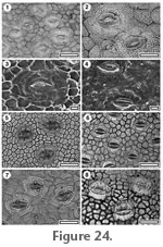
Identification. Identification of Rhizophoraceae is based on the large, cyclocytic stomatal complexes, which have a trend towards alignment, and which are placed within isodiametric epidermal cells which are distinctly smaller than the dimensions of the stomatal complex (see also
Ramassamy and Kannabiran 1996,
Farooqui 2001). The family Rhizophoraceae includes both terrestrial and mangrove species, but this combination of epidermal characters (Figure 24.5-24.7) appears to be unique to Bruguiera, Ceriops, and Rhizophora which are closely related genera within the mangrove tribe Rhizophoreae within the Rhizophoraceae (Setoguchi et al. 1999). Pelliciera (Pellicieraceae) a mangrove in the tropical American region has the aligned stomatal complexes and small epidermal cells, but the stomatal complex morphology is distinctly different (Figure 24.8). Other Rhizophoraceae species available in the reference collection have very different epidermal morphologies. CUT-Z-JAG is therefore regarded as a mangrove of the family Rhizophoraceae.
 Reference specimen and locality: SB0694, R-20. Reference specimen and locality: SB0694, R-20.
Referred specimens and occurrence: SB0693, R-20.
Stomatal complexes. Stomatal distribution over leaf surfaces unknown, stomata evenly spread, isolated, randomly oriented, unclear, bimodal size range with occasional giant stomata. Subsidiary cells (3–4) irregularly shaped, (generally not possible to count under TLM because of surface ornamentation), periclinal walls same thickness as normal epidermal cells, ornamentation continuous with that of epidermal cells. Guard cell pair outline circular, outer margin obscured under TLM by surface ornamentation, length 28–45 µm, (medium-large), with prominent polar rods. Outer stomatal ledge elliptical, thicker than normal epidermal cells, extending from outer edge of stoma, pore elliptical.
Epidermal cells. Epidermal cell flanges not distinct under TLM because of surface ornamentation, normal cells highly variable from isodiametric to elongate, approximately the same size as the stomata, anticlinal walls curved to sinuous, unbuttressed, ornamented with 'flowing' pattern of many fine ridges.
Indumentum. Trichome insertion scars sparse, with slightly thickened poral rims.
Distinguishing features. Having subsidiary and epidermal cells entirely covered by flowing fine ridges.
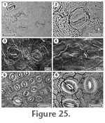
Identification. Ilex cornuta (Aquifoliaceae) has a similar ornamentation of fine, flowing ridges, and has giant stomata (Figure 25.5-25.6). However, its epidermal cell flanges are distinct, despite the ornamentation, and the subsidiary cells are smaller and more adpressed to the guard cells.
 Reference specimen and locality: SB0395, R-75. Reference specimen and locality: SB0395, R-75.
Referred specimens and occurrence: SB0737, R-12.
Stomatal complexes. Stomatal distribution over leaf surfaces unknown, stomata evenly spread, isolated, randomly oriented, unclear, size range bimodal, with distinct 'giant stomata' present, (distinguished by being much narrower and longer and by having bands of fine ridges flowing away from them). Subsidiary cells difficult to count under TLM because of ornamentation of short, cuspate ridges which are concentric about the stoma, periclinal walls same thickness as normal epidermal cells, ornamented with 3–4 ridges parallel with, and on either side of the stomata. Guard cell pair outline elliptical, outlined by a well-defined anticlinal wall, clearly separated by polar walls, length 23-38 µm. Outer stomatal ledge elliptical, extending from outer edge of stoma, same thickness as normal epidermal cells, pore elliptical.
Epidermal Cells. Epidermal cell flanges clearly visible using TLM, normal cells highly variable from isodiametric to elongate, approximately the same size as the stomata, anticlinal walls curved, unbuttressed, ornamented with discontinuous, cuspate, thick ridges.
Indumentum. Glabrous.
Distinguishing features. Having a prominent ornamentation of discontinuous, cuspate ridges.
Identification. The extant Nemopanthus mucronata (Aquifoliaceae,
Figure 26.4) has a similar ornamentation of short, cuspate ridges, as well as having 'giant stomata' which are longer and narrower than normal stomata and have ridges flowing away from them. Identification is suggested to be with Aquifoliaceae.
 Reference specimen and locality: SB0335, R-74. Reference specimen and locality: SB0335, R-74.
Referred specimens and occurrence: SL5326, R-06; SL5254, R-13; SB0720, R-21; SL5164, R-33; SB0334, R-74.
Stomatal complexes. Stomatal distribution over leaf surfaces unknown, stomata evenly spread, isolated, randomly oriented, anisocytic, size range unimodal. Subsidiary cells (2–4) irregularly shaped, periclinal walls same thickness as normal epidermal cells, ornamented with thick ridges concentric about the stomata. Guard cell pair outline elliptical, outlined by a well-defined anticlinal wall, with prominent T-piece thickenings at polar ends, length 40–55 µm (large). Outer stomatal ledge elliptical, same thickness as normal epidermal cells, extending from outer edge of stoma, pore elliptical.
Epidermal Cells. Epidermal cell flanges clearly visible using TLM, normal cells highly variable from isodiametric to elongate, typically as long as, although narrower than typical epidermal cells, anticlinal walls curved, unbuttressed, unornamented.
Indumentum. Glabrous.
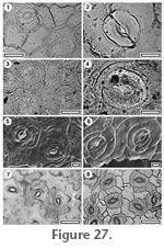
Distinguishing features. Large anisocytic stomatal complexes with an ornamentation of concentric ridges restricted to subsidiary cells.
Identification. Large, anisocytic stomatal complexes with an ornamentation of relatively thick ridges over the subsidiary cells suggests Aquifoliaceae (Figure 27.7-27.8). Finer ridges would be more indicative of Myrsinaceae.
Key to Miscellaneous angiosperm cuticle
1. Cuticle has distinct papillae. Group A.
1. Cuticle does not have distinct papillae. 2.
2. Cuticle has distinct surface striations. Group B.
2. Cuticle does not have distinct striations. 3.
3. Stomatal orientation is aligned. Group C.
3. Stomatal orientation is random. Group D.
Group A.
Cuticle with distinct papillae
1. Papillae ridged, No trichomes or trichome scars present. CUT-Z-JBB
1. Papillae not ridged. 2.
2. Papillae over whole epidermis. CUT-Z-GDA
2. Papillae distinct only around stomata. 3.
3. Papillae smooth, discrete, large, multicellular trichome insertion scars present. CUT-Z-GCF
3. Papillae broad, cuticle glabrous. CUT-Z-JAD
Group B.
Cuticle with prominent striations or grooves.
1. Cuticle very thin, epidermal cell outlines not clear. CUT-Z JJE
1. Epidermal cell outlines clear under TLM. 2.
2. Striations confined to, or most prominent in area around stomata. 3.
2. Striations spread over whole cuticle. 4.
3. Striations thick, forming two or three very discontinuous rings around stomata, cuticle glabrous. CUT-Z-JCF
3. Striations thin and many, cuticle with many persistent trichome bases. CUT-Z-JCC
4. Epidermal cells sinuous. CUT-Z-JJA
4. Epidermal cells polygonal or slightly wavy. CUT-Z-JJB
Group C.
Stomatal orientation is aligned.
1. Thick, simple trichome bases present. CUT-Z-JEB
1. No trichome bases. 2.
2. Epidermal cell outlines obscured by granularity of cuticle. CUT-Z-JEJ
2. Epidermal cell outlines clear. CUT-Z-JEA
Group D.
Cuticle without papillae, striations or aligned stomatal complexes.
1. Stomatal complex paracytic. CUT-Z-JCA
1. Stomatal complex not paracytic. 2.
2. Stomatal complex anisocytic, largest subsidiary cell in complex much larger than guard cell pair. 3.
2. Stomatal complex not anisocytic. 4.
3. Epidermal and subsidiary cell walls straight. CUT-Z-JJF
3. Epidermal and subsidiary cell walls wavy. CUT-Z-JEF
4. Epidermal cells sinuous. 5.
4. Epidermal cells not sinuous.6.
5. Trichome bases common, fine venation reflected in shape of epidermal cells. CUT-Z-JCJ
5. Trichome bases essentially absent, fine venation not reflected in shape of epidermal cells. CUT-Z-JJH
6. Stomatal aperture surrounded by a massively thickened ring. CUT-Z-JDH
6. Stomatal aperture not surrounded by a massively thickened ring. 7.
7. Stomata without prominent outer stomatal ledge. 8.
7. Stomata with very prominent outer stomatal ledge. 10.
8. Stomatal complexes infrequent, surrounded by a double ring of subsidiary cells. CUT-Z-JAA
8. Stomatal complexes common, not surrounded by a double ring of subsidiary cells. 9.
9. Stomata round, subsidiary cell arrangement highly variable, their cuticle of same thickness as normal epidermal cells. CUT-Z-JJI
9. Stomata elliptical, typically four subsidiary cells disorganised, thinner than normal epidermal cells. CUT-Z-JAE
10. Stomatal complexes staurocytic, stomatal pore slit-like. CUT-Z-JEE
10. Stomatal complexes not staurocytic, stomatal pore elliptical or sub-circular 11.
12. Epidermal cells isodiametric, small poral trichome bases present. CUT-Z-JED
12. Epidermal cells irregular to elongate, glabrous. CUT-Z-JBH
 Reference specimen and locality: SB0378, R-75. Reference specimen and locality: SB0378, R-75.
Referred specimens and occurrence: SL5208, R-08; SB0377, R-75; SB0710, R-76, SL5371, R-102.
Stomatal complexes. Stomatal distribution in clear areolar groups, isolated, randomly oriented, cyclocytic or anomocytic, size range unimodal, length about 5-7 µm (small).
Epidermal cells. Flanges often indistinct under TLM (because of ornamentation of papillae), isodiametric, walls straight. From the centre of each normal epidermal and sometimes on venal epidermal cells, projects a single, longitudinally ridged papilla, which expands distally either slightly, or to cover the entire epidermal cell.
Indumentum. Glabrous.
Distinguishing features. Having longitudinally ridged papilla, which expand slightly, distally.
Identification. This cuticle is regarded as the same as that published by
Hill (1991) as Eucryphia. Barnes and Jordan (2000) did not believe Hill's (1991) identification of the fossils to Eucryphia was correct. Hill described E. microstoma as brachyparacytic (a feature of extant Eucryphia) although this is not apparent in his figures.
 Reference specimen and locality: SL5433, R-102. Reference specimen and locality: SL5433, R-102.
Stomatal complexes. Stomatal distribution unknown, isolated, randomly oriented, brachyparacytic, size range unimodal, length 20–22 µm (medium). Subsidiary cells not papillate, not obscured by papillae.
Epidermal cells. Flanges often indistinct under TLM (because of ornamentation of papillae), isodiametric, walls straight. Surface of each normal epidermal cell projecting outwards to form a single, smooth papilla.
Indumentum. With persistent, simple trichomes (staining much darker than normal epidermal cuticle under TLM), surrounded by epidermal cells radially elongated, and either papillae-free or with subdued papillae, forming foot-cells.
Distinguishing features. Having papillae and persistent simple trichomes.
Reference specimen and locality: SB0371, R-75.
Stomatal complexes. Stomatal distribution over leaf surfaces unknown, stomata in clear areolar areas between veins, isolated, randomly oriented, size range probably bimodal. Each stomata is surrounded by a ring of papillae (4-6) projecting from the subsidiary cells, one from each cell. Normal stomata completely obscured by papillae, but larger ones have the guard cells visible, typical length about 13 µm (small) but up to about 25 µm (medium).
Epidermal cells. Epidermal cell flanges clearly visible using TLM, normal cells isodiametric, approximately the same size as the stomata, anticlinal walls curved, unbuttressed, unornamented but papillate, each cell with a single smooth papilla formed from most of the cell surface. Epidermal cells over fine veins distinguished by being more rectangular and not papillate.
Indumentum. Trichome insertion scars common (trichomes deciduous), situated over veins, consisting of a sub-circular opening with a smooth; Slightly thickened rim, 25–45 µm in diameter, leading to a chamber flanked by many cells with walls thicker than typical venal epidermal cells.
Distinguishing features. Having large papillate epidermal cells and distinctive compound trichome insertion pits.
Reference specimen and locality: SB0725, R-23.
Stomatal complexes. Stomatal distribution over leaf surfaces unknown, stomata evenly spread. isolated, randomly oriented, size range unimodal, length around 25 µm (medium). Pore massively thickened, slit-like. Subsidiary cells 4-5, periclinal walls raised up as bubble-like papillae, anticlinal walls thicker than normal epidermal cells.
Epidermal cells. Isodiametric, rounded, granular, raised up slightly as papillae, but less than the subsidiary cells.
Indumentum. Glabrous.
Distinguishing features. CUT-Z-JAD is similar to CUT-L-DCG with regard to the massive thickening around the stomatal pore and subsidiary cells. However, it is distinct on the basis of papillae and higher number of subsidiary cells.
 Reference specimen and locality: SB0732, R-6. Reference specimen and locality: SB0732, R-6.
Referred specimens and occurrence: SB0704, R-12; SB0734, R-22.
Stomatal complexes. Stomatal distribution over leaf surfaces unknown, stomata evenly spread, isolated, randomly oriented, size range unimodal. Subsidiary cell number and shape unclear, periclinal walls same thickness as normal epidermal cells, ornamented with fine ridges concentric about the stomata. Guard cell pair outline elliptical, not outlined by a clear anticlinal wall, length 23–40 µm, (medium-large). Outer stomatal ledge elliptical, thicker than normal epidermal cells, extending over inner edge of stoma, pore narrowly elliptic.
Epidermal cells. Epidermal cell periclinal and anticlinal walls very thin (anticlinal walls of epidermal cells not clear under TLM), unornamented.
Indumentum. With sparse trichome insertion scars (trichomes deciduous and therefore trichome type unknown), inserted between epidermal cells, with a massively thickened poral rim and radiating walls.
Distinguishing features. Very thin cuticle, but with massively thickened poral trichome insertion scars.
Reference specimen and locality: SB0727, R-6.
Stomatal complexes. Stomatal distribution over leaf surfaces unknown, stomata evenly spread, isolated, randomly oriented, size range unimodal. Subsidiary cells not distinguished under TLM and therefore construction unclear, periclinal walls same thickness as normal epidermal cells, ornamented with a more or less continuous ridge immediately external to the outer stomatal ledge, and further out by 2–3 very discontinuous ridges concentric about the stomata. Guard cell pair outline elliptical, length 23–40 µm (medium-large). Outer stomatal ledge elliptical, same thickness as normal epidermal cells, extending from outer edge of stoma, pore elliptical.
Epidermal cells. Epidermal cell flanges somewhat diffuse, normal cells highly variable from isodiametric to elongate, approximately the same size as the stomata, anticlinal walls curved, unbuttressed, unornamented.
Distinguishing features. Having 2–3 very discontinuous ridges concentric about the stomata.
Indumentum. Glabrous.
 Reference specimen and locality: SB0741, R-30. Reference specimen and locality: SB0741, R-30.
Stomatal complexes. Stomatal distribution over leaf surfaces unknown, stomata evenly spread, isolated, brachyparacytic, size range unimodal, Subsidiary cells irregularly shaped, periclinal walls same thickness as normal epidermal cells, ornamented with fine ridges concentric about the stomata. Outer margin obscured under TLM by surface ornamentation, length 25–30 µm (medium). Outer stomatal ledge elliptical, same thickness as normal epidermal cells, extending from outer edge of stoma, pore elliptical.
Epidermal cells. Epidermal cell flanges clearly visible using TLM, normal cells highly variable from isodiametric to elongate, approximately the same size as the stomata, anticlinal walls straight, unbuttressed, unornamented.
Indumentum. Hirsute, with abundant, simple, persistent trichomes inserted between epidermal cells. Epidermal cells around trichome base unmodified.
Distinguishing features. Brachyparacytic stomatal complexes and persistent trichomes.
Reference specimen and locality: SB0723, R-7.
Referred specimens and occurrence: SB0723, R-7; SL5320, R-06; SL5206, R-07; SB0765, R-22; SL5373, R-102.
Stomatal complexes. Stomatal distribution over leaf surfaces unknown, isolated, randomly oriented, size range unimodal. Subsidiary cells (4–6) irregularly-shaped, difficult to distinguish by shape from epidermal cells, but with more granular inner cuticular surface, construction variable from actinocytic to possibly only one subsidiary cell, periclinal walls as thick as or often distinctly thinner than normal epidermal cells, ornamented by many fine ridges radiating from or flowing around the stomata. Guard cell pair outline elliptical, outlined by a well-defined anticlinal wall, length 15–28 µm (medium), clearly separated by polar walls. Outer stomatal ledge elliptical, same thickness as normal epidermal cells, extending from outer edge of stoma, pore elliptical.
Epidermal cells. Epidermal cell flanges clearly visible using TLM, normal cells highly variable from isodiametric to elongate, distinctly larger than the stomata, anticlinal walls sinuous, unbuttressed, ornamented with 'flowing' pattern of many fine ridges.
Indumentum. Glabrous.
Distinguishing features. Elliptical stomata, sinuous epidermal cells and an ornamentation of fine flowing ridges.
 Reference specimen and locality: SB0753, R-76. Reference specimen and locality: SB0753, R-76.
Stomatal complexes. Stomatal distribution over leaf surfaces unknown, stomata evenly spread, isolated, showing a clear trend towards alignment, unclear, size range unimodal. Subsidiary cells difficult to count under TLM, periclinal walls same thickness as normal epidermal cells, unornamented. Guard cell pair outline elliptical, outlined by a well-defined anticlinal wall, length 30–45 µm, (medium-large), some wall development between guard cells, and sometimes development of T-piece thickenings at the poles. Outer stomatal ledge elliptical, same thickness as normal epidermal cells, extending from outer edge of stoma, pore elliptical.
Epidermal Cells. Epidermal cell flanges clearly visible using TLM, normal cells elongated, approximately the same size as the stomata, anticlinal walls straight, unbuttressed, unornamented.
Indumentum. With trichome insertion scars (trichomes deciduous). Trichome scars sparse, inserted between epidermal cells. Epidermal cells around trichome scar mostly radially elongate, often modified by tangential divisions to form an irregular sub-circular zone of foot cells, poral rim massively thickened and frill-like.
Distinguishing features. Having a massively thickened and frill-like trichome poral rim.
Reference specimen and locality: SB0726, R-22.
Stomatal complexes. Stomatal distribution over leaf surfaces unknown, stomata evenly spread, isolated, showing a clear trend towards alignment, construction unclear, size range unimodal. Subsidiary cells not possible to count under TLM because of surface ornamentation, periclinal walls same thickness as normal epidermal cells, unornamented. Guard cell pair outline elliptical, not outlined by a clear anticlinal wall, length 18–35 µm (medium), with prominent T-piece thickenings at polar ends. Outer stomatal ledge elliptical, extending from outer edge of stoma, thicker than normal epidermal cells, pore elliptical.
Epidermal cells. Epidermal cell flanges not distinct under TLM because of surface ornamentation, unornamented.
Indumentum. Glabrous.
Distinguishing features. Elliptical stomata with a clear trend towards alignment and prominent T-pieces.
Reference specimen and locality: SB0717, R-12.
Referred specimens and occurrence: SL5322, R-06; SB0717, R-12; SL5245, R-13; SL5047, R-21; SL5181, R-38; SL4998, R-47; SL5266, R-76.
Stomatal complexes. Stomatal distribution over leaf surfaces unknown, stomata evenly spread, isolated (but very rare networking noted), showing a clear trend towards alignment, anomocytic, size range unimodal. Subsidiary cells (4–5) irregularly shaped, periclinal walls same thickness as normal epidermal cells, unornamented. Guard cell pair outline circular, outlined by a very well-defined anticlinal wall, length 30–43 µm (medium-large), clearly separated by polar walls. Outer stomatal ledge sub circular, extending over centre of stoma, thicker than over normal epidermal cells, but surrounded by a very narrow thin zone, pore broad, sub-circular.
Epidermal cells. Epidermal cell flanges clearly visible using TLM, normal cells highly variable from isodiametric to elongate, approximately the same size, or slightly smaller than the stomata, anticlinal walls straight to curved, unbuttressed, unornamented.
Indumentum. Glabrous.
Distinguishing features. Circular stomata showing a clear trend towards alignment.
 Reference specimen and locality: SB0764, R-21. Reference specimen and locality: SB0764, R-21.
Referred specimens and occurrence: SB0762, R-25.
Stomatal complexes. Stomatal distribution over leaf surfaces unknown, stomata evenly spread, isolated, randomly oriented, brachyparacytic, size range unimodal. Subsidiary cells typically elongate tangential to stomata, often longer than the stomata, unequally sized, periclinal walls same thickness as normal epidermal cells, unornamented. Guard cell pair outline elliptical, outlined by a well-defined anticlinal wall, length 18–35 µm (medium), clearly separated by polar walls. Outer stomatal ledge elliptical, extending over inner edge of stoma, thicker than normal epidermal cells, pore narrowly elliptic.
Epidermal cells. Epidermal cell flanges clearly visible using TLM, normal cells highly variable from isodiametric to elongate, approximately the same size as the stomata, anticlinal walls sinuous, unbuttressed, unornamented.
Indumentum. Glabrous.
Distinguishing features. Brachyparacytic stomatal complexes, sinuous anticlinal walls and glabrous.
 Reference specimen and locality: SB0739, R-9. Reference specimen and locality: SB0739, R-9.
Referred specimens and occurrence:
Stomatal complexes. Stomatal distribution over leaf surfaces unknown, stomata evenly spread, isolated, randomly oriented, anisocytic (largest subsidiary cell is three to four times larger than the stoma), size range unimodal. Subsidiary cell periclinal walls same thickness as normal epidermal cells, unornamented. Guard cell pair outline circular, outlined by a well-defined anticlinal wall, length 15–20 µm (medium), little polar development between guard cells (guard cells appear as continuous ring). Outer stomatal ledge sub circular, extending from outer edge of stoma, same thickness as normal epidermal cells, pore elliptical.
Epidermal cells. Epidermal cell flanges clearly visible using TLM, normal cells highly variable from isodiametric to elongate, from approximately the same size as the stomata to distinctly larger, anticlinal walls straight, unbuttressed, unornamented.
Indumentum. Glabrous.
Distinguishing features. Anisocytic stomatal complexes in which the largest subsidiary cell is three to four times larger than the stomata and with straight epidermal cell anticlinal walls. Differs from CUT-Z-JEF by having straight rather than sinuous walls.
Reference specimen and locality: SL5426, R-34; SB0698, R-76.
Stomatal complexes. Stomatal distribution over leaf surfaces unknown, stomata evenly spread, isolated, randomly oriented, anisocytic (largest cell is three-four times larger than the stoma), size range unimodal. Subsidiary cell periclinal walls same thickness as normal epidermal cells, unornamented. Guard cell pair outline circular, outlined by a well-defined anticlinal wall, length 20–25 µm (medium), little polar development between guard cells (guard cells appear as continuous ring). Outer stomatal ledge sub circular, extending from outer edge of stoma, same thickness as normal epidermal cells, pore elliptical.
Epidermal cells. Epidermal cell flanges clearly visible using TLM, normal cells elongated, from approximately the same size as the stomata to distinctly larger, anticlinal walls sinuous, unbuttressed, unornamented.
Indumentum. Glabrous.
Distinguishing features. Anisocytic stomatal complexes in which the largest subsidiary cell is three to four times larger than the stomata and with sinuous epidermal cell anticlinal walls. Differs from CUT-Z-JJF by having sinuous rather than straight walls.
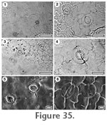 Reference specimen and locality: SB0763, R-102. Reference specimen and locality: SB0763, R-102.
Stomatal complexes. Stomatal distribution over leaf surfaces unknown, stomata evenly spread, often networked, randomly oriented, anomocytic, size range unimodal. Subsidiary cell periclinal walls same thickness as normal epidermal cells, unornamented. Guard cell pair outline circular, outlined by a well-defined anticlinal wall, length 15–28 µm (medium), clearly separated by polar walls. Outer stomatal ledge sub circular, extending over centre of stoma, thicker than normal epidermal cells, pore elliptical - sub circular.
Epidermal cells. Epidermal cell flanges clearly visible using TLM, though cuticle generally thin, normal cells elongated, approximately the same size as the stomata, anticlinal walls sinuous, unbuttressed, unornamented.
Indumentum. With trichome insertion scars. Trichome scars common (trichome deciduous), inserted between epidermal cells. Epidermal cells around trichome scar modified into radially-elongate foot-cells (6–8), and thickened to form a poral rim and radial walls.
Distinguishing features. Circular stomata with sinuous epidermal cells and common simple trichome insertion scars. Differs from CUT-Z-JJH by the presence of trichome insertion scars.
Reference specimen and locality: SB0701, R-26.
Referred specimens and occurrence: SL5346, R-11; SL5055, R-21; SB0748, R-23; SL5088, R-25; SB0701, R-26; SL5336, R-50; SL5329, R-75.
Stomatal complexes. Stomatal distribution over leaf surfaces unknown, stomata evenly spread, isolated, randomly oriented, anomocytic, size range unimodal. Subsidiary cell periclinal walls same thickness as normal epidermal cells, unornamented. Guard cell pair outline circular, outlined by a well-defined anticlinal wall, length 28–35 µm (medium), some wall development between guard cells, and sometimes development of T-piece thickenings at the poles. Outer stomatal ledge sub circular, extending from outer edge of stoma, thicker than normal epidermal cells, pore elliptical - sub circular.
Epidermal cells. Epidermal cell flanges clearly visible using TLM, normal cells highly variable from isodiametric to elongate, from approximately the same size as the stomata to distinctly larger, anticlinal walls sinuous, unbuttressed, unornamented.
Indumentum. Glabrous.
Distinguishing features. Circular stomata with sinuous epidermal cells and absence of trichome insertion scars. Differs from CUT-Z-JCJ by the absence of trichome insertion scars.
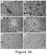 Reference specimen and locality: SB0712, R-25. Reference specimen and locality: SB0712, R-25.
Stomatal complexes. Stomatal distribution over leaf surfaces unknown, stomata evenly spread, isolated, randomly oriented, unclear, size range unimodal. Subsidiary cell periclinal walls same thickness as normal epidermal cells, ornamented with a very broad, massive, and lumpy peristomatal rim. Guard cell pair outline difficult to distinguish, outer margin obscured under TLM by surface ornamentation, length 25–28 µm (medium). Outer stomatal ledge elliptical, extending from outer edge of stoma, same thickness as normal epidermal cells, pore elliptical.
Epidermal cells. Epidermal cell flanges clearly visible using TLM, normal cells isodiametric, distinctly smaller than the stomata, anticlinal walls straight, unbuttressed, unornamented.
Indumentum. With trichome insertion scars (trichomes deciduous). About 7 ft cells surround scar; slightly radially elongated.
Distinguishing features. Having a very broad, massive and lumpy peristomatal rim.
Reference specimen and locality: SB0350, R-74.
Stomatal complexes. Stomatal distribution over leaf surfaces unknown, stomata evenly spread, isolated, randomly oriented, cyclocytic, appears to be two rings of subsidiary cells, size range unimodal. Subsidiary cell periclinal walls thinner than over normal epidermal cells, unornamented. Guard cell pair outline elliptical, not outlined by a clear anticlinal wall, length 25–38 µm (medium), little polar development between guard cells (guard cells appear as continuous ring). Outer stomatal ledge not clear, thinner than normal epidermal cells, pore elliptical.
Epidermal cells. Epidermal cell flanges clearly visible using TLM but of varying thickness, normal cells isodiametric, approximately the same size, or slightly smaller than the stomata, anticlinal walls straight, unbuttressed, unornamented.
Indumentum. Glabrous.
Distinguishing features. Sparse stomatal complexes with apparently two rings of subsidiary cells.
Reference specimen and locality: SB0754, R-76.
Stomatal complexes. Stomatal distribution over leaf surfaces unknown, stomata in areoles, sometimes networked, randomly oriented, size range unimodal, construction very variable from apparently anomocytic to having one or two subsidiary cells (distinguished by being narrower than normal epidermal cells). Subsidiary cell periclinal walls same thickness as normal epidermal cells, unornamented. Guard cell pair outline elliptical, outlined by a well-defined anticlinal wall, length 15–20 µm (medium), some wall development between guard cells, and sometimes development of T-piece thickenings at the poles. Outer stomatal ledge elliptical, extending from outer edge of stoma, same thickness as normal epidermal cells, pore elliptical.
Epidermal cells. Epidermal cell flanges clearly visible using TLM, though cuticle generally thin, normal cells highly variable from isodiametric to elongate, approximately the same size as the stomata, cells over both major and fine venation more elongate, anticlinal walls curved, unbuttressed, unornamented.
Indumentum. Glabrous.
Distinguishing features. Thin cuticle with thin outer stomatal ledges and stomatal complexes varying from anomocytic to having one or two subsidiary cells.
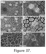 Reference specimen and locality: SL5071, R-21. Reference specimen and locality: SL5071, R-21.
Referred specimens and occurrence: SL5201, R-06; SL5217, R-11; SL5225, R-12; SL5253, R-13; SL5066, R-21; SB0718, R-26; SL5161, R-33; SL5182, R-39; SL5001, R-47; SB0345, R-74; SB0374, R-75.
Stomatal complexes. Stomatal distribution over leaf surfaces unknown, stomata evenly spread, isolated, randomly oriented, cyclocytic, size range unimodal. Subsidiary cells (4–5) with periclinal walls as thick as, or sometimes slightly thicker than normal epidermal cells, but also with a distinctly thinner zone around the outer stomatal ledge, unornamented. Guard cell pair outline elliptical, not outlined by a clear anticlinal wall, length 15–25 µm (medium), some wall development between guard cells, and sometimes development of T-piece thickenings at the poles. Outer stomatal ledge elliptical, extending from outer edge of stoma, thinner than normal epidermal cells, pore elliptical.
Epidermal cells. Epidermal cell flanges clearly visible using TLM, normal cells highly variable from isodiametric to elongate, from approximately the same size as the stomata to distinctly larger, anticlinal walls thick, straight to curved, unbuttressed, unornamented and with a particularly smooth texture.
Indumentum. With trichome insertion scars (trichomes deciduous). Trichome scars sparse, inserted between epidermal cells. Epidermal cells around trichome scar modified by tangential divisions to form a sub-circular zone of foot cells (5–6), and thickened to form a poral rim and radial walls.
Distinguishing features. Cyclocytic stomatal complexes and epidermal cells with a particularly smooth texture
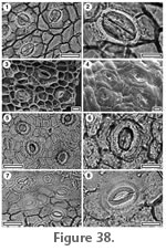 Reference specimen and locality: SB0738, R-29. Reference specimen and locality: SB0738, R-29.
Referred specimens and occurrence: SB0342, R-74; SB0375, R-75.
Stomatal complexes. Stomatal distribution over leaf surfaces unknown, stomata evenly spread, isolated, randomly oriented, staurocytic-cyclocytic, size range unimodal. Subsidiary cells (4–5) isodiametric-elongate tangential to stoma, periclinal walls same thicker than normal epidermal cells, unornamented. Guard cell pair outline difficult to see under TLM, length about 38–50 µm (medium to mostly large). Outer stomatal ledge prominent, sub circular; Slightly pinched at the poles, extending from within the outer edge of stoma, thinner than normal epidermal cells but somewhat granular, and surrounded by a prominent ring of thin (not granular) cuticle, pore slit-like.
Epidermal cells. Epidermal cell flanges clearly visible using TLM (very thick), normal cells isodiametric-elongate, typically similar size to stomata, anticlinal walls straight, unbuttressed, unornamented.
Indumentum. Glabrous.
Distinguishing features. Mostly large staurocytic-cyclocytic stomatal complexes.
Reference specimen and locality: SB0724, R-24.
Referred specimens and occurrence: SL5302, R-105.
Stomatal complexes. Stomatal distribution over leaf surfaces unknown, stomata evenly spread, isolated, randomly oriented, anomocytic, size range unimodal. Subsidiary cells (5–6) isodiametric, periclinal walls same thickness as normal epidermal cells, unornamented. Guard cell pair outline elliptical to circular, length 33–45 µm (medium-large). Outer stomatal ledge sub circular, extending from outer edge of stoma, thicker than normal epidermal cells, pore elliptical - sub circular.
Epidermal cells. Epidermal cell flanges clearly visible using TLM (very thick), normal cells isodiametric, typically slightly smaller than the stomata, anticlinal walls straight, unbuttressed, unornamented.
Indumentum. With trichome insertion scars. Trichome scars common (trichomes deciduous), inserted between epidermal cells, diameter much smaller than normal epidermal cell. Epidermal cells around trichome scar modified slightly into radially elongate foot-cells (6–7), with no distinct thickening of walls.
Distinguishing features. Prominent outer stomatal ledges, and isodiametric epidermal cells, trichome insertion scars with a diameter much smaller than epidermal cells.
Reference specimen and locality: SB0337, R-74.
Stomatal complexes. Stomatal distribution over leaf surfaces unknown, stomata evenly spread, isolated, randomly oriented, construction unclear, size range unimodal. Subsidiary cell periclinal walls same thickness as normal epidermal cells, unornamented. Guard cell pair outline elliptical, outlined by an indistinct anticlinal wall, length 25–35 µm (medium), with prominent T-piece thickenings at polar ends, same thickness as normal epidermal cells, Outer stomatal ledge extending from outer edge of stoma, pore elliptical.
Indumentum. Glabrous.
Epidermal cells. Epidermal cell flanges clearly visible using TLM, normal cells highly variable from isodiametric to elongate, approximately the same size as the stomata, anticlinal walls curved, unbuttressed, unornamented.
Distinguishing features. Bland morphology which has prominent T-piece thickenings of the stomata and lack of any distinct stomatal construction.
|