|
|
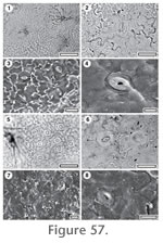
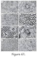
APPENDIX 3 (continued).TAXA OF UNCERTAIN FAMILYGroup 1. Papillae presentKey to papillate cuticle1. Cuticle hisute (trichomes persistent). 2. 1. Trichomes deciduous. 3. 2. Papillae simple and smooth. CUT-Z-ECH 2. Papillae peltate. CUT-Z-ABI 3. Stomatal complexes scattered.4. 3. Stomatal complexes in clearly defined patches. CUT-Z-ABD 4. Papillae restricted to subsidiary cells CUT-Z-ABA 4. Papillae on both subsidiary and normal epidermal cells. 5. 5. Papillae much smaller than the epidermal cell. CUT-Z-ABC 5. Papillae comparable in size to the epidermal cell. CUT-Z-ABB
CUT-Z-ABA
Referred specimens and occurrence: SL0281, BL-01; SL0240, BL-04; SL1279, BL-05; SL0365, BL-09; SL3088, BL-28; SL3178, BL-33; SL2097, GL-07; SL2140, GL-08; SL2219, GL-12; SL2895, GL-20; SL2904, GL-21; SL2929, GL-22; SL3109, GL-24; SL2965, GL-25; SL0383, Mata-01; SB0845, Mata-06. Stomatal complexes. Stomatal complexes in areoles, isolated, randomly oriented, laterocyclic, size range unimodal. Subsidiary cells (9–11) isodiametric, periclinal walls of same thickness as normal epidermal cells, granular. With a single papilla which projects horizontally over the stomatal pore. Unornamented. Guard cell pair outline difficult or impossible to see in TLM view, sunken below subsidiary cells and epidermal cells, outer margin obscured under TLM by surface topography, length 20–35 µ (medium). Epidermal Cells. Epidermal cell flanges clearly visible using TLM, normal cells isodiametric (cells over major veins more elongate), approximately the same size as the stomata, anticlinal walls curved to wavy, unbuttressed, inner surfaces very slightly granular, outer surfaces smooth, unornamented, occasionally papillate. Indumentum. Attachment scars of deciduous trichomes common, scattered over venal and non-venal regions but slightly more common over veins, inserted between (7–9) epidermal cells modified only slightly, with a thickened rim. Scar diameter much smaller than a normal epidermal cell. Smooth papillae present. Distinguishing features. Distinguished from CUT-Z-ABC in that papillae are restricted to subsidiary cells.
CUT-Z-ABC Reference Specimen and locality: SB1334, BL-32. Referred specimens and occurrence: SL0084, BL-08; SL1182, GL-01; SL2654, GL-04; SL2090, GL-07; SL1760, Sthd-012. Stomatal complexes. Stomatal complexes in areoles, isolated, randomly oriented, cyclocytic, size range bimodal, with distinct 'giant stomatal complexes' present (restricted to venal regions). Subsidiary cells (6–10) isodiametric, periclinal walls of same thickness as normal epidermal cells, smooth. With a single papilla which projects horizontally over the stomatal pore. Unornamented. Guard cell pair outline difficult or impossible to see in TLM view, sunken below subsidiary cells, outer margin obscured under TLM by surface topography, length c. 20 µ (medium). Epidermal Cells. Epidermal cell flanges clearly visible using TLM, normal cells isodiametric (cells over both major and fine venation more elongate), approximately the same size as the stomata, anticlinal walls curved to wavy, unbuttressed, inner surfaces slightly granular, outer surfaces slightly rough, unornamented. Indumentum. Attachment scars of deciduous trichomes common, restricted to regions over veins, inserted between (6–11) epidermal cells modified into radially elongate foot cells, insertion 'peg' diameter much smaller than a normal epidermal cell, but the trichome then expands abruptly above the insertion. Papillae in patches, generally restricted to regions surrounding stomatal complexes, one papilla per cell, formed by the projection of a discrete region within the boundaries of the epidermal cell, smooth, much smaller than the epidermal cell, oriented vertically. Distinguishing features. Distinguished from CUT-Z-ABB by the smaller papillae – much smaller than the epidermal cells, and from CUT-Z-ABA in that papillae are found on subsidiary cells and commonly also on some normal epidermal cells.
CUT-Z-ABI
Referred specimens and occurrence: SL3167, BL-33. Stomatal complexes. Stomatal complexes apparently in well-defined areoles. All details of stomata obscured under TLM by surface ornamentation and trichomes. Epidermal Cells. Details of normal epidermal cells obscured under TLM because of surface thickenings, but cells over some veins are distinguished by being unornamented. Indumentum. Hirsute and with dense peltate extensions of epidermal cells. Trichomes abundant, uniseriate, apparently more common over venal areas.
CUT-Z-ECH Reference Specimen and locality: SL1208, GL-01. Referred specimens and occurrence: SL3156, GL-01; SB1208, Mata-01. Stomatal complexes. Stomatal complexes in areoles, isolated, number of subsidiary cells unclear because of obscuring papillae, size range bimodal, with distinct 'giant stomatal complexes' present. Subsidiary cells (4–6) isodiametric, periclinal walls of same thickness as normal epidermal cells, smooth, each with a single papilla which projects horizontally over the stomatal pore. Guard cell pair outline difficult or impossible to see in TLM view as obscured by surface topography, but appears to be sunken with respect to subsidiary and epidermal cells. Epidermal Cells. Epidermal cell flanges clearly visible using TLM, normal cells isodiametric (cells over both major and fine venation more elongate), approximately the same size as the stomata, anticlinal walls straight, unbuttressed, inner surfaces smooth, outer surfaces rough, unornamented. Indumentum. Hirsute and papillate, trichomes common, scattered over venal and non-venal regions, simple, inserted between (6-11) unmodified or sometimes radially elongate epidermal cells, insertion peg diameter very small, but trichome base similar in size to a normal epidermal cell. The periclinal surface of each epidermal cells projects upwards as a single smooth papilla. Distinguishing features. Distinguished from other taxa with persistent trichomes in that the entire surface of all epidermal and subsidiary cells are raised up as papillae.
CUT-Z-ABB
Referred specimens and occurrence: SL1277, BL-05; SL3081, BL-28; SL2141, GL-08; SL2173, GL-09; SL2180, GL-10; SL2205, GL-11; SL2218, GL-12; SL2849, GL-18; SL2890, GL-20; SL2943, GL-23; SL3101, GL-24; SL2968, GL-25; SL2975, GL-26. Stomatal complexes. Stomatal distribution over leaf surfaces unknown, randomly oriented, actinocytic, size range bimodal, with distinct 'giant stomatal complexes' present. Subsidiary cells (4–6) isodiametric, periclinal walls of same thickness as normal epidermal cells; smooth, each with a single papilla that projects horizontally over the stomatal pore. Guard cell pair outline difficult or impossible to see in TLM view as obscured by surface topography. Epidermal Cells. Epidermal cell flanges clearly visible using TLM but only away from stomatal areas; anticlinal walls curved to wavy (cells over major veins more elongate); approximately the same size as the stomata; unbuttressed; inner and outer surfaces moderately granular, unornamented. Indumentum. Scars of deciduous trichomes and papillae present. Trichome attachment scars abundant, scattered over venal and non-venal regions, inserted between (6–9) epidermal cells modified into radially elongate foot cells, scar diameter much smaller than a normal epidermal cell. Papillae in patches, generally restricted to regions surrounding stomatal complexes, with one smooth, discrete papilla per cell, comparable in size to the epidermal cel. Distinguishing features. Distinguished from CUT-Z-ABC by the larger papillae – comparable in size to the epidermal cell.
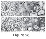
CUT-Z-ABD Reference Specimen and locality: SB1332, Mata-03. Referred specimens and occurrence: SL0286, BL-01; SL0238, BL-04; SL1275, BL-05; SL2697, BL-06; SL0026, BL-07; SL0098, BL-08; SL2467, BL-09; SL2250, BL-14; SL2271, BL-16; SL2292, BL-18; SL3052, BL-24; SL3076, BL-27; SL3083, BL-28; SL3093, BL-29; SL1224, BL-30; SL2517, BL-31; SL1377, BL-32; SL2537, BL-33; SL1179, GL-01; SL1187, GL-02; SL2658, GL-04; SL2190, GL-10; SL2201, GL-11; SL2216, GL-12; SL2823, GL-16; SL2838, GL-17; SL2861, GL-18; SL2883, GL-20; SL2899, GL-21; SL2918, GL-22; SL2939, GL-23; SL3104, GL-24; SL2949, GL-25; SL2993, GL-27; SL3018, GL-29; SL1057, Mata-01; SL0334, Mata-23. Stomatal complexes. Stomatal complexes in areoles, isolated, randomly oriented, cyclocytic, size range unimodal. Subsidiary cells (4–6) isodiametric, periclinal walls of same thickness as normal epidermal cells; with peltate scales. Unornamented. Guard cell pair outline distinctly narrow and almost rectangular, but difficult or impossible to see in TLM view; overarched by subsidiary cells, outer margin obscured under TLM by surface topography; length c. 10 µ (small). Epidermal Cells. Epidermal cell flanges clearly visible using TLM; normal cells highly variable from isodiametric to elongate (cells over both major and fine venation more elongate); distinctly larger than the stomata; anticlinal walls curved to wavy; unbuttressed; inner surfaces coarsely granular, outer surfaces smooth, ornamented with short ridges perpendicular or parallel to some stomatal complexes. Indumentum. Attachment scars of deciduous trichomes common, restricted to regions over veins, inserted between (9–11) epidermal cells modified by tangential divisions to form a sub-circular zone of foot cells, scar diameter similar in size to a normal epidermal cell. Distinguishing features. Distinguished by having stomatal complexes in clearly defined patches and which are entirely hidden below peltate cuticular extensions. Group 2. Stomatal complexes generally alignedKey to cuticle with Stomatal complexes generally aligned1. Outer stomatal ledge elliptical, Guard cell pair outline difficult or impossible to see in TLM view. CUT-Z-AAE 1. Outer stomatal ledge sub circular, guard cell pair outline ovate. CUT-Z-CGJ
CUT-Z-AAE
Referred specimens and occurrence: SL0122, BL-03. Stomatal complexes. Stomatal complexes in areoles, isolated, showing a clear trend towards alignment, cyclocytic, size range unimodal. Subsidiary cells (5–7) irregularly-shaped, difficult to count under TLM, periclinal walls of same thickness as normal epidermal cells, smooth, unornamented. Guard cell pair outline difficult or impossible to see in TLM view, not outlined by a clear anticlinal wall, at same level as subsidiary cells, length 17–21 µ (medium). Outer stomatal ledge elliptical, same thickness as normal epidermal cells, extending over inner edge of stoma. Pore narrowly elliptic. Epidermal Cells. Epidermal cell flanges clearly visible using TLM, normal cells isodiametric (cells over both major and fine venation more elongate), distinctly smaller than the stomata, anticlinal walls straight, unbuttressed, very smoothly textured, unornamented. Indumentum. Glabrous.
CUT-Z-CGJ Reference Specimen and locality: SL0352, BL-05. Stomatal complexes. Stomatal complexes evenly spread, isolated, with a tendency to be aligned with one another at least in patches, anomocytic, size range bimodal, with distinct 'giant stomatal complexes' present. Subsidiary cells (4–5) irregularly-shaped, periclinal walls of same thickness as normal epidermal cells, smooth, unornamented. Guard cell pair outline ovate, outlined by a well-defined anticlinal wall, length 27–30 µ (medium), at same level as subsidiary cells (exposed on surface), some wall development between guard cells, and sometimes development of T-piece thickenings at the poles. Outer stomatal ledge sub circular, same thickness as normal epidermal cells, extending over whole stoma, with a broad, sub-circular pore. Epidermal Cells. Epidermal cell flanges clearly visible using TLM, normal cells highly variable from isodiametric to elongate (cells over major veins more elongate), approximately the same size as the stomata, anticlinal walls straight, unbuttressed, slightly rough, unornamented. Indumentum. Glabrous. Distinguishing features. Glabrous cuticle with virtually naked kidney-shaped guard cells. Group 3. Epidermal cells highly sinuousKey to cuticle with highly sinuous epidermal cells1. Guard cell pair outline circular, clearly visible under TLM. CUT-Z-AAC 1. Guard cell pair outline ovate, but obscured by the strong outer stomatal ledge. CUT-Z-AAD
CUT-Z-AAC
Referred specimens and occurrence: SL2597, BL-01; SL2284, BL-18; SL1183, GL-01; SL1198, GL-02; SL2675, GL-04; SL2187, GL-10; SL2200, GL-11; SL2831, GL-17; SL3012, GL-28. Stomatal complexes. Stomatal complexes evenly spread, isolated, randomly oriented, cyclocytic, size range unimodal. Subsidiary cells (8–10, difficult to count under TLM) isodiametric; periclinal walls of same thickness as normal epidermal cells; smooth; unornamented. Guard cell pair outline circular; outlined by a well-defined anticlinal wall; length 25–30 µ (medium); at same level as subsidiary cells (exposed on surface); with prominent polar rods. Outer stomatal ledge sub circular, thinner than normal epidermal cells, extending over whole stoma, with a broad, sub-circular pore. Epidermal Cells. Epidermal cell flanges clearly visible using TLM; normal cells irregular (cells over both major and fine venation elongate); approximately the same size as the stomata; anticlinal walls markedly sinuous; strongly buttressed; inner surfaces slightly granular, outer surfaces smooth, unornamented. Indumentum. Attachment cars of deciduous trichomes sparse, restricted to regions over veins, simple, inserted between (6–9) epidermal cells modified by tangential divisions to form a sub-circular zone of foot cells, scar diameter much smaller than a normal epidermal cell. Distinguishing features. Distinguished from CUT-Z-AAD in having very round stomatal complexes and many subsidiary cells.
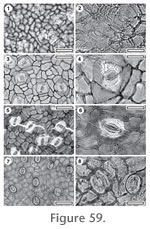
CUT-Z-AAD Reference Specimen and locality: SB1291, Mata-03. Referred specimens and occurrence: SL3295, BL-04; SL2207, GL-12; SL2842, GL-18; SL3123, Sthd-198. Stomatal complexes. Stomatal complexes evenly spread, isolated, randomly oriented, laterocyclic, or cyclocytic, developmentally tangenticytic, size range unimodal. Subsidiary cells (4–5) typically elongate tangential to the stoma, periclinal walls of same thickness as normal epidermal cells, smooth, ornamented with groups of ridges radiating from the stoma. Guard cell pair outline ovate, but obscured by the strong outer stomatal ledge, at same level as subsidiary cells (exposed on surface), length 32–38 µ (medium). Outer stomatal ledge sub circular, same thickness as normal epidermal cells, extending over inner edge of stoma, with an elliptical pore. Epidermal Cells. Epidermal cell flanges clearly visible using TLM, normal cells highly variable from isodiametric to elongate (cells over major veins more elongate), approximately the same size as the stomata, anticlinal walls markedly sinuous, strongly buttressed, smoothly textured ornamented with ridges of irregular course. Indumentum. Attachment scars of deciduous trichomes sparse, not noted over veins, inserted between (7–8) epidermal cells modified by tangential divisions to form a sub-circular zone of foot cells, scar diameter much smaller than a normal epidermal cell. Distinguishing features. Distinguished from CUT-Z-AAC in having ovate stomatal complexes and fewer (about 4) subsidiary cells. Group 4. Stomatal complexes surrounded by a prominent, broad peristomatal ringKey to cuticle with prominent peristomal ring1. Peristomal ring margins sharply defined, epidermal cells distinctly small than stoma. CUT-Z-ADD 1. Peristomal ring margins smooth, epidermal cells of comparable size to the stoma. 2. 2. Outer stomatal ledge separated from the peristomatal rim by a narrow groove. Stoma with prominent thickenings of polar rods. CUT-Z-EIG 2. Outer stomatal ledge separated from the peristomatal rim by a broad groove. Stoma without thickenings of polar rods. CUT-Z-FGA
CUT-Z-ADD
Referred specimens and occurrence: SL3137, GL-01. Stomatal complexes. Stomatal complexes evenly spread, isolated, randomly oriented, cyclocytic, all some, or none of the subsidiary cells have been modified by a tangential division (developmentally stephanocytic bicyclic), size range bimodal, with distinct 'giant stomatal complexes' present (separated from other stomatal complexes by a distinct stomatal-free zone). Subsidiary cells (3–5) typically elongate tangential to stoma, periclinal walls of same thickness as normal epidermal cells, granular, ornamented with a thick, sharply defined and slightly lumpy peristomatal rim. Guard cell pair outline ovate, outer margin obscured under TLM by surface topography, length c. 50 µ (large). Outer stomatal ledge sub circular, same thickness as normal epidermal cells, extending over whole stoma, separated from the peristomatal rim by a deep, narrow groove. Pore broad, sub-circular. Epidermal Cells. Epidermal cell flanges clearly visible using TLM, normal cells isodiametric (cells over veins not distinguished by shape), distinctly smaller than the stomata, anticlinal walls curved to wavy, unbuttressed, inner surfaces slightly granular, outer surfaces smooth, unornamented. Indumentum. Glabrous. Distinguishing features. Distinguished by the massive peristomatal ring.
CUT-Z-EIG Reference Specimen and locality: SL1764, Sthd-011. Stomatal complexes. Stomatal complexes evenly spread, isolated, randomly oriented, cyclocytic, developmentally tangenticytic, size range unimodal. Subsidiary cells (4–5) isodiametric, periclinal walls of same thickness as normal epidermal cells, smooth, ornamented with a thick, smooth-edged peristomatal rim. Guard cell pair outline ovate, outlined by a well-defined anticlinal wall, at same level as subsidiary cells (exposed on surface), length 42–43 µ (large), with prominent thickenings of polar rods. Outer stomatal ledge sub circular, thinner than normal epidermal cells, extending over whole stoma, with a broad, sub-circular pore, separated from the peristomatal rim by a narrow groove. Epidermal Cells. Epidermal cell flanges clearly visible using TLM, normal cells isodiametric (cells over veins not distinguished by shape), approximately the same size as the stomata, anticlinal walls curved to wavy, or markedly sinuous, unbuttressed, smoothly textured, unornamented. Indumentum. Glabrous.
CUT-Z-FGA Reference Specimen and locality: SL2016, Sthd-010. Stomatal complexes. Stomatal complexes isolated, possibly with some general alignment although this is not clear, number of subsidiary cells unclear, size range unimodal. Periclinal walls of same thickness as normal epidermal cells, smooth, ornamented with a thick, smooth edged peristomatal rim. Guard cell pair outline difficult or impossible to see in TLM view, outer margin obscured under TLM by surface ornamentation, length 35–41 µ (large-medium). Outer stomatal ledge sub circular, thicker than normal epidermal cells, extending over whole stoma, separated from peristomatal rim by a broad, shallow groove. Pore broad, sub-circular. Epidermal Cells. Epidermal cell flanges somewhat diffuse, normal cells elongate (unclear if cells over veins are distinguished), approximately the same size as the stomata, anticlinal walls curved to wavy, unbuttressed, slightly granular, flanges reflected on the surface by discontinuous, broad ridges, otherwise unornamented . Indumentum. Glabrous. Group 5. Persistent trichomes presentKey to cuticle with persistent trichomes1. Trichome width multicellular at base. CUT-Z-ABG 1. Trichome width single-celled at base. 2. 2. Cuticle surface ornamented with ridges. 3. 2. Cuticle surface unornamented. 4. 3. Normal epidermal cells curved to wavy and unbuttressed. CUT-Z-CDC 3. Normal epidermal cells sinuous and buttressed. CUT-Z-FFE 4. Stomatal complexes not anisocytic. 5. 4. Stomatal complexes often anisocytic. 6. 5. Stomatal complexes anomocytic (developmentally tangeticytic), Stomatal pore elliptical. CUT-Z-ACJ 5. Stomatal complexes cyclocytic, developmentally stephanocytic Stomatal pore with a key-hole outline. CUT-Z-ABH 6. Cuticle over the stoma so thin it often breaks away, or, under TLM view, gives impression of the outer stomatal ledge is "floating". CUT-Z-EEJ 6. Cuticle over the stoma typically robust, not breaking. 7. 7. Stomatal outline unclear under TLM, no T-thickenings at poles. CUT-Z-FGB 7. Stomatal outline clear under TLM, often T-thickenings at poles. CUT-Z-EDI
CUT-Z-FGB
Stomatal complexes. Stomatal complexes evenly spread, isolated, randomly oriented, anisocytic, size range unimodal. Subsidiary cells (3) with periclinal walls thinner than that over normal epidermal cells, smooth, ornamented with ridges concentric about the stoma. Guard cell pair outline difficult or impossible to see in TLM view, not outlined by a clear anticlinal wall, at same level as subsidiary cells (exposed on surface), length 20–22 µ (medium), little polar development between guard cells (guard cells appear as continuous ring). Outer stomatal ledge elliptical, thinner than normal epidermal cells, only extending over inner edge of stoma, with a narrowly elliptic pore. Epidermal Cells. Epidermal cell flanges clearly visible using TLM, normal cells highly variable from isodiametric to elongate (cells over veins not distinguished by shape), approximately the same size as the stomata, anticlinal walls curved to wavy, unbuttressed, smoothly textured, unornamented. Indumentum. Hirsute, trichomes common, scattered over venal and non-venal regions, uniseriate, a chain of 4-5 individual cells, inserted between radially elongate epidermal cells. Insertion diameter much smaller than a normal epidermal cell, but then the trichome base expanded abruptly above the insertion point. Distinguishing features. Clearly distinguished by the uniseriate trichomes which are inserted over several modified epidermal cells.
CUT-Z-EDI Reference Specimen and locality: SL2002, Sthd-072. Stomatal complexes. Stomatal complexes evenly spread, isolated, randomly oriented, anisocytic, or unevenly paracytic, size range unimodal. Subsidiary cells (2–4) irregularly-shaped, periclinal walls of same thickness as normal epidermal cells, smooth, unornamented. Guard cell pair outline circular, not outlined by a clear anticlinal wall, at same level as subsidiary cells (exposed on surface), length 22–29 µ (medium), little wall development between guard cells but often with prominent T-thickenings at poles. Outer stomatal ledge sub circular, thinner than normal epidermal cells, extending over whole stoma, with a broad, sub-circular pore. Epidermal Cells. Epidermal cell flanges clearly visible using TLM, normal cells isodiametric (cells over major veins more elongate), approximately the same size as the stomata, anticlinal walls curved to wavy, buttressed, inner and outer surfaces smooth, unornamented. Indumentum. Bases of trichomes common, probably restricted to regions over veins (unclear), persistent, but all cases the main length of the trichome has broken off irregularly just above the base, inserted between unmodified epidermal cells, basal diameter much larger than a normal epidermal cell.
CUT-Z-CDC
Stomatal complexes. Stomatal complexes evenly spread, isolated, randomly oriented, number of subsidiary cells unclear (difficult to count under TLM), size range bimodal, with distinct 'giant stomatal complexes' present. Subsidiary cell periclinal walls thicker than over normal epidermal cells, smooth, ornamented with many ridges which tend to flow around the stoma but also frequently cross portions of the stoma. Guard cell pair outline ovate, outer margin obscured under TLM by surface ornamentation, length 11–12 µ (small), at same level as subsidiary cells (exposed on surface). Outer stomatal ledge sub circular, thinner than normal epidermal cells, extending over whole stoma, with a broad, sub-circular pore. Epidermal Cells. Epidermal cell flanges poorly visible under TLM and only visible away from stomatal complexes, normal cells highly variable from isodiametric to elongate (cells over major veins more elongate), anticlinal walls curved to wavy, unbuttressed; inner and outer surfaces smooth, ornamented with bands of irregular ridges parallel to stomatal complexes and radiating from trichomes. Distinguishing features. Distinguished from other taxa with persistent trichomes in that the surface is densely ornamented with irregular ridges concentric or roughly parallel to stomatal complexes and which radiate from trichomes.Indumentum. Trichomes sparse; scattered over venal and non-venal regions, simple, mostly broken of irregularly above the base, trichome diameter similar in size to a normal epidermal cell. Epidermal cells around trichome base (6–7) modified by tangential divisions to form 4–5 rings of foot cells.
CUT-Z-FFE Reference Specimen and locality: SL2120, GL-07. Stomatal complexes. Stomatal complexes evenly spread, isolated, randomly oriented, number of subsidiary cells unclear, possibly brachyparacytic, size range unimodal. Periclinal walls of same thickness as normal epidermal cells, smooth; ornamented with bands of ridges projecting at right angles to the stomatal axis. Guard cell pair outline difficult or impossible to see in TLM view because of ornamentation; not outlined by a clear anticlinal wall; at same level as subsidiary cells (exposed on surface), length 17–18 µ (medium). Outer stomatal ledge elliptical, same thickness as normal epidermal cells, extending over whole stoma, with a narrowly elliptic pore. Epidermal Cells. Epidermal cell flanges clearly visible using TLM; normal cells highly variable from isodiametric to elongate (cells over veins not distinguished by shape); approximately the same size as the stomata; anticlinal walls markedly sinuous; buttressed; smoothly textured, ornamented with ridges of irregular course. Indumentum. Trichomes common; uniseriate although some may branch from the base, or are perhaps in clusters, trichome diameter similar in size to a normal epidermal cell.
CUT-Z-EEJ Reference Specimen and locality: SL1778, Sthd-030. Referred specimens and occurrence: SL1731, Sthd-017. Stomatal complexes. Stomatal complexes evenly spread, isolated, randomly oriented, anisocytic, size range unimodal. Subsidiary cells (3–4) irregularly-shaped, (hard to count as radial flanges of subsidiary cells are not well developed), periclinal walls of same thickness as normal epidermal cells, smooth, unornamented. Guard cell pair outline ovate, not outlined by a clear anticlinal wall, at same level as subsidiary cells (exposed on surface), length 18–23 µ (medium), little polar development between guard cells (guard cells appear as continuous ring). Outer stomatal ledge elliptical, much thinner than normal epidermal cells (often broken away), extending over whole stoma. Pore narrowly elliptic. Epidermal Cells. Epidermal cell flanges clearly visible using TLM, normal cells isodiametric (cells over veins not distinguished by shape), approximately the same size as the stomata, anticlinal walls straight, unbuttressed, smoothly textured, unornamented. Indumentum. Bases of trichomes common, apparently uniseriate although most broken off in the lower segment, inserted between (6-8) epidermal cells modified into radially elongate foot cells, but with no distinct thickening of walls, basal diameter similar to much larger than a normal epidermal cell. Distinguishing features. Distinguished from other taxa with persistent trichomes in the very thin cuticle over the stoma which often breaks away, or gives impression of the outer stomatal ledge "floating" in a TLM view.
CUT-Z-ABG
Referred specimens and occurrence: SL3131, GL-02. Stomatal complexes. Stomatal complexes evenly spread, isolated, randomly oriented, anisocytic, size range unimodal. Subsidiary cells (3–4, difficult to count under TLM) irregularly-shaped, periclinal walls of same thickness as normal epidermal cells, smooth, ornamented with ridges concentric about the stoma. Guard cell pair outline circular, outlined by a well-defined anticlinal wall, at same level as subsidiary cells (exposed on surface), length 12–16 µ (small). Outer stomatal ledge sub circular, thinner than normal epidermal cells, extending over whole stoma, with a broad, sub-circular pore. Epidermal Cells. Epidermal cell flanges clearly visible using TLM, normal cells highly variable from isodiametric to elongate (cells over veins not distinguished by shape), approximately the same size as the stomata, anticlinal walls markedly sinuous, unbuttressed, inner surface very finely granular, outer surface smooth, ornamented with ridges of irregular course. Indumentum. Trichomes abundant, scattered over venal and non-venal regions, multicellular, unbranched, lower part of trichome formed of several elongate cells packed side by side, which then narrows to a single apical cell, base diameter equivalent to 3–6 epidermal cells. Distinguishing features. Distinguished from other taxa with persistent trichomes in that the trichomes are multicellular.
CUT-Z-ACJ Reference Specimen and locality: SB1350, BL-32. Stomatal complexes. Stomatal complexes evenly spread, isolated, randomly oriented, anomocytic (developmentally tangeticytic), size range unimodal. Subsidiary cells (4–5) irregularly-shaped, periclinal walls of same thickness as normal epidermal cells, smooth, unornamented. Outlined by a well-defined anticlinal wall, at same level as subsidiary cells (exposed on surface), length 13–15 µ (small). Outer stomatal ledge elliptical, same thickness as normal epidermal cells, extending over inner edge of stoma, with an elliptical pore. Epidermal Cells. Epidermal cell flanges clearly visible using TLM, normal cells highly variable from isodiametric to elongate (cells over at least some veins are more elongate), approximately the same size as the stomata, anticlinal walls markedly sinuous, unbuttressed, smoothly textured, unornamented. Indumentum. Hirsute, trichomes common, probably restricted to regions over veins (unclear), uniseriate, 3–5 cells long, the cells nearest the trichome base are sometimes about as long as they are wide, more distal cells are 5–6 times as long as wide, total length about 88–113 m, the base is a circular, non-poral cell, but the base may expand over surrounding epidermal cells, basal diameter of similar or distinctly larger size than normal epidermal cells. Epidermal cells around trichome base unmodified
CUT-Z-ABH
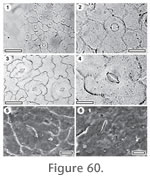
Referred specimens and occurrence: SL2101, GL-07; SL2160, GL-09. Stomatal complexes. Stomatal complexes evenly spread, isolated, randomly oriented, cyclocytic, developmentally stephanocytic bicyclic, size range unimodal (but with wide range of stomatal size). Subsidiary cells (5–7) typically elongate tangential to stoma, with periclinal walls thinner than that over normal epidermal cells, smooth, unornamented. Guard cell pair outline circular, outer margin obscured under TLM by robustness of outer stomatal ledge, at same level as subsidiary cells (stomatal complexes sunken so that outer stomatal ledges at same level as surface of epidermal cells), length 20–28 µ (medium). Outer stomatal ledge sub circular, same thickness as normal epidermal cells, extending over whole stoma. Pore broad, but narrows abruptly towards the poles (a key-hole outline) and may be slightly off-set from one guard cell to the other. Epidermal Cells. Epidermal cell flanges clearly visible using TLM, normal cells isodiametric (cells over major veins more elongate), distinctly larger than the stomata, anticlinal walls curved to wavy, unbuttressed, inner surfaces slightly rough, outer surfaces smooth, unornamented. Indumentum. Trichomes common, scattered over venal and non-venal regions, persistent, inserted between (6–12) epidermal cells modified into radially elongate foot cells, with no distinct thickening of walls, diameter of insertion 'peg' much smaller than a normal epidermal cell, but then the trichome expands abruptly to about the same size or larger than a typical epidermal cell. Distinguishing features. CUT-Z-ABH is distinguished from CUT-Z-AJD in the presence of trichomes. It is distinguished from other taxa with persistent trichomes in that the subsidiary cell periclinal walls are much thinner than either normal epidermal cells or the outer stomatal ledges. Group 6. Stomatal complexes paracyticKey to cuticle with paracytic stomatal complexes1. Stomatal complexes large, prominent T-piece thickening at guard cell poles, epidermal cell anticlinal walls sinuous. CUT-Z-CDD 1. Stomatal complexes medium, slight T-piece thickening at guard cell poles, epidermal cell anticlinal walls wavy to slightly sinuous. CUT-Z-CFF
CUT-Z-CCD
Referred specimens and occurrence: SL0234, BL-04; SL3075, BL-27; SL3152, GL-01; SL4791, GL-07; SL2128, GL-08; SL2870, GL-20; SL2920, GL-22; SL2947, GL-25; SL1235, BL-32. Stomatal complexes. Stomatal complexes evenly spread, isolated, randomly oriented, brachyparacytic, size range unimodal. Subsidiary cells more or less equal to unevenly sized, typically expanding to longer than the guard cells, periclinal walls of same thickness as normal epidermal cells, smooth, unornamented. Guard cell pair outline elongate, often with flattened poles, outlined by a well-defined anticlinal wall, length 38–40 µ (large), at same level as subsidiary cells (exposed on surface), with very prominent T-piece thickenings at polar ends. Outer stomatal ledge sub circular, thicker than normal epidermal cells, extending over centre of stoma, with a broad, sub-circular pore. Epidermal Cells. Epidermal cell flanges clearly visible using TLM, normal cells elongate (cells over major veins more elongate), distinctly larger than the stomata, anticlinal walls markedly sinuous, unbuttressed, inner surfaces granular, outer surfaces smooth, unornamented. Indumentum. Attachment scars of deciduous trichomes rare, inserted between (c. 6) epidermal cells modified by tangential divisions to form a sub-circular zone of foot cells, scar diameter much smaller than a normal epidermal cell. Distinguishing features. Distinguished by the large and unevenly sized brachyparacytic subsidiary cells.
CUT-Z-CFF Reference Specimen and locality: SL0359, BL-05. Referred specimens and occurrence: SL3181, BL-01; SL2541, GL-02. Stomatal complexes. Stomatal complexes evenly spread, isolated, randomly oriented, brachyparacytic, or anisocytic, size range unimodal. Subsidiary cells (2–4) sometimes forming distinct polar cells (right angles to stomatal orientation) and 2 lateral cells (parallel to stomatal orientation), periclinal walls of same thickness as normal epidermal cells, smooth, unornamented. Guard cell pair outline ovate, outlined by a well-defined anticlinal wall, length 22–30 µ (medium), at same level as subsidiary cells (exposed on surface), with slight T-piece thickenings at polar ends. Outer stomatal ledge sub circular, thinner than normal epidermal cells, extending over whole stoma, with a broad, sub-circular pore. Epidermal Cells. Epidermal cell flanges clearly visible using TLM, normal cells highly variable from isodiametric to elongate (cells over veins not distinguished by shape), distinctly larger than the stomata, anticlinal walls wavy to slightly sinuous, unbuttressed, outer surfaces smooth, but with periclinal walls irregularly raised leaving grooves over the anticlinal walls. Indumentum. Glabrous. Distinguishing features. Although the typical subsidiary cell pattern of CUT-Z-CFF is brachyparacytic, the underlying stomatal pattern is probably anisocytic. A spiralling process of cell division results in two subsidiary cells of reasonably equal size and on opposite sides of the guard cells. A third subsidiary can sometimes be distinguished. This taxon thus differs fundamentally from truly brachyparacytic taxa. Group 7. Stomatal complexes anisocyticKey to cuticle with anisocytic stomatal complexes1. Trichomes present and persistent. CUT-Z-AJH 1. Cuticle glabrous. 2. 2. Epidermal cell periclinal walls distinctly raised, leaving very narrow, deep grooves over anticlinal walls. CUT-Z-ECE 2. Epidermal cell periclinal walls not distinctly raised. 3. 3. Epidermal cell anticlinal walls sinuous, prominent polar rod thickenings. CUT-Z-JII 3. Epidermal cell anticlinal walls straight to wavy, without prominent polar rod thickenings. 4. 4. Subsidiary cells granular, stomatal complex with distinctive angular outline. CUT-Z-FFB 4. Subsidiary cells not granular, stomatal complex without a distinctive angular outline. 5. 5. Peristomatal rim present. CUT-Z-FJG 5. Peristomatal rim absent. 6. 6. Normal epidermal cells elongate, outer stomatal ledge distinctly thinner than normal epidermal cells, subsidiary cells (3–4). CUT-Z-FCD 6. Normal epidermal cells isodiametric to elongate, outer stomatal ledge slightly thinner than normal epidermal cells, subsidiary cells (4–5). CUT-Z-EIH
CUT-Z-AJH
Referred specimens and occurrence: SL0087, BL-08; SL1196, GL-02. Stomatal complexes. Stomatal complexes evenly spread, isolated, randomly oriented, anisocytic, heliocyctic, size range bimodal, with distinct 'giant stomatal complexes' present. Subsidiary cells (3) of varying size, curved and generally tangential to the guard cells, periclinal walls of same thickness as normal epidermal cells; smooth; unornamented. Guard cell pair outline circular; outlined by a well-defined anticlinal wall; length 13–15 µ (small); at same level as subsidiary cells (exposed on surface); with prominent T-piece thickenings at polar ends. Outer stomatal ledge sub circular, same thickness as normal epidermal cells, extending over whole stoma, with a broad, sub-circular pore. Epidermal Cells. Epidermal cell flanges clearly visible using TLM; normal cells elongate (cells over major veins more elongate); distinctly larger than the stomata; anticlinal walls curved to wavy; unbuttressed; smoothly textured, unornamented. Indumentum. Hirsute; trichome bases common; scattered over venal and non-venal regions but slightly more common over veins, persistent, but all cases broken off near base, inserted between unmodified epidermal cells, basal diameter similar in size to a normal epidermal cell. Distinguishing features. Distinguished from CUT-Z-JII in the presence of persistent trichomes.
CUT-Z-FCD Reference Specimen and locality: SL3016, GL-28. Stomatal complexes. Stomatal complexes in areoles, isolated, randomly oriented, anisocytic, (this appears to be the basic state, but there are often more than the definitive three subsidiary cells), size range unimodal. Subsidiary cells (3–4) irregularly-shaped, periclinal walls of same thickness as normal epidermal cells, smooth, unornamented. Guard cell pair outline ovate, outlined by a well-defined anticlinal wall, at same level as subsidiary cells (exposed on surface), length 22–30 µ (medium), with prominent T-piece thickenings at polar ends. Outer stomatal ledge sub circular, distinctly thinner than normal epidermal cells, extending over centre of stoma, with a broad, sub-circular pore. Epidermal Cells. Epidermal cell flanges clearly visible using TLM, normal cells elongate (cells over major veins more elongate), approximately the same size as the stomata, anticlinal walls curved to wavy, unbuttressed, smoothly textured, unornamented. Indumentum. Glabrous.
CUT-Z-JII Reference Specimen and locality: SB1356, BL-32. Stomatal complexes. Stomatal complexes in areoles, sometimes networked, randomly oriented, anisocytic, size range unimodal. Subsidiary cells (3–4) with periclinal walls of same thickness as normal epidermal cells, inner surfaces granular, unornamented. Guard cell pair outline circular, outlined by a well-defined anticlinal wall, at same level as subsidiary cells (exposed on surface), length 16–20 µ (medium), with very prominent polar rods. Outer stomatal ledge sub circular, same thickness as normal epidermal cells, extending over whole stoma, with a broad, sub-circular pore. Epidermal Cells. Epidermal cell flanges clearly visible using TLM, normal cells elongate (cells over major veins more elongate), approximately the same size as the stomata, anticlinal walls markedly sinuous, slightly buttressed, inner and outer surfaces smooth, unornamented. Indumentum. Glabrous. Distinguishing features. Distinguished from CUT-Z-AJH in being glabrous and having buttressed epidermal cell walls.
CUT-Z-FFB
Stomatal complexes. Stomatal complexes in areoles, isolated, randomly oriented, size range unimodal. Subsidiary cells (3–4) with periclinal walls of same thickness as normal epidermal cells, granular, unornamented, with angular outline. Guard cell pair outline elongate, outlined by a well-defined anticlinal wall, at same level as subsidiary cells (though may be slightly sunken below surface), length 22–30 µ (medium). Outer stomatal ledge same thickness as normal epidermal cells, with a narrowly elliptic pore. Epidermal Cells. Epidermal cell flanges clearly visible using TLM, normal cells isodiametric (cells over both major and fine venation more elongate), approximately the same size as the stomata, anticlinal walls straight, unbuttressed, inner surfaces smooth, outer surfaces rough, unornamented. Indumentum. Glabrous. Identification. The distinctive angular outline to the stomatal complex is reminiscent of the Oxylobium illicifolium illustrated by Jordan (1997). This may indicate relationships with the Fabaceae.
CUT-Z-FJG Reference Specimen and locality: SL2506, BL-03. Referred specimens and occurrence: SL2238, GL-12. Stomatal complexes. Stomatal complexes in areoles, isolated, randomly oriented, anisocytic, heliocyctic, size range unimodal. Subsidiary cells (3–5) with periclinal walls of same thickness as normal epidermal cells, smooth, ornamented with a thick peristomatal rim. Guard cell pair outline elongate, outer margin obscured under TLM by surface topography, length 21–30 µ (medium). Outer stomatal ledge elliptical, same thickness as normal epidermal cells, extending over whole stoma, with a narrowly elliptic pore. Epidermal Cells. Epidermal cell flanges clearly visible using TLM, normal cells highly variable from isodiametric to elongate (cells over veins not distinguished by shape), approximately the same size as the stomata, anticlinal walls straight, unbuttressed, smoothly textured, unornamented. Indumentum. Glabrous. Distinguishing features. Stomatal complex is elongate with rounded ends and with a peristomatal ridge. CUT-Z-CDE differs in having many subsidiary cells and no peristomatal ridge. Identification. The distinctive angular outline to the stomatal complex is reminiscent of the Oxylobium ilicifolium (Andrews) Domin illustrated by Jordan (1997). This may indicate relationships within the Fabaceae.
CUT-Z-ECE
Referred specimens and occurrence: SL2686, Sthd-090. Stomatal complexes. Stomatal complexes evenly spread, isolated, randomly oriented, anisocytic, heliocyctic, size range unimodal. Subsidiary cells (3) of varying size, curved and generally tangential to the guard cells, periclinal walls of same thickness as normal epidermal cells, smooth, ornamented with a single, irregular rim. Guard cell pair outline circular, outlined by a well-defined anticlinal wall, at same level as subsidiary cells (exposed on surface), length 25–35 µ (medium). Outer stomatal ledge sub circular, same thickness as normal epidermal cells, extending over whole stoma, with a broad, sub-circular pore. Epidermal Cells. Epidermal cell flanges not clearly visible under TLM because of surface thickenings, normal cells very irregular or elongate (cells over veins not distinguished by shape), approximately the same size as the stomata, anticlinal walls curved to wavy, very thick, unbuttressed, smoothly textured, periclinal walls raised, leaving very narrow, deep grooves over anticlinal walls. Indumentum. Glabrous. Distinguishing features. Distinguished from other taxa with anisocytic stomatal complexes in having anticlinal walls of epidermal and subsidiary cells reflected by strong ridges on cuticle surface. The subsidiary and epidermal cell anticlinal walls are also markedly depressed below general surface of cuticle.
CUT-Z-EIH Reference Specimen and locality: SL1566, Sthd-004. Stomatal complexes. Stomatal complexes evenly spread, isolated, randomly oriented, anisocytic, all some, or none of the subsidiary cells may have been modified by a tangential division, size range bimodal, with distinct 'giant stomatal complexes' present (separated from other stomatal complexes by a distinct stomatal-free zone). Subsidiary cells (4–5) irregularly-shaped, difficult to count under TLM, periclinal walls of same thickness as normal epidermal cells, smooth, unornamented. Guard cell pair outline circular, not outlined by a clear anticlinal wall, at same level as subsidiary cells (exposed on surface), length 25–32 µ (medium), some wall development between guard cells, and sometimes development of T-piece thickenings at the poles. Outer stomatal ledge sub circular, slightly thinner than normal epidermal cells, extending over whole stoma, with a broad, sub-circular pore. Epidermal Cells. Epidermal cell flanges clearly visible using TLM, normal cells isodiametric to elongate (cells over veins not distinguished by shape), anticlinal walls curved to wavy, unbuttressed, smoothly textured, unornamented. Indumentum. Glabrous. Group 8. Multi Cellular trichome bases presentKey to cuticle with multi-cellular trichome bases1. Trichome attachment scar with thickened, raised rim, cells forming base of scar not thickened. CUT-Z-ECC 1. Trichome attachment scar without thickened, raised rim, cells below scar distinctly thickened. CUT-Z-EID
CUT-Z-ECC
Referred specimens and occurrence: SL1351, BL-05; SL1249, BL-32; SB1308, Mata-03; SL1751, Sthd-016; SL1704, Sthd-019; SL1871, Sthd-078; SL1527, Sthd-111. Stomatal complexes. Stomatal distribution over leaf surfaces hypostomatic, stomatal complexes tightly clustered in areoles, frequently networked, randomly oriented, anisocytic, size range bimodal, with distinct 'giant stomatal complexes' present. Subsidiary cells (3) isodiametric, periclinal walls thicker than over normal epidermal cells, smooth, ornamented with many ridges which tend to flow around the stoma. Guard cell pair outline ovate, outlined by a well-defined anticlinal wall, length 9–16 µ (small), at same level as subsidiary cells (exposed on surface), little polar development between guard cells (guard cells appear as continuous ring). Outer stomatal ledge sub circular, thinner than normal epidermal cells, extending over whole stoma, with a broad, sub-circular pore. Epidermal Cells. Epidermal cell flanges clearly visible using TLM, normal cells isodiametric (cells over major veins vaguely or inconsistently more elongate), approximately the same size as the stomata, anticlinal walls straight, unbuttressed, inner and outer surfaces smooth, ornamented with many fine ridges over subsidiary cells, running parallel to stomatal axis. Indumentum. Insertion scars of deciduous trichomes abundant, scattered over venal and non-venal regions but slightly more common over veins, inserted over several (3–7) modified epidermal cells. Scar diameter similar in size to a normal epidermal cell. Epidermal cells are selectively thickened to form a slightly raised, thickened rim, although the cells within the rim are not thickened. Distinguishing features. Distinct in having stomatal complexes with well-defined (by thickness of anticlinal walls) subsidiary cells which occur in tight clusters, within which there is some networking, and by the many and prominent trichome attachment scars which are situated over several epidermal cells.
CUT-Z-EID Reference Specimen and locality: SL1718, Sthd-018. Referred specimens and occurrence: SL3176, BL-33. Stomatal complexes. Stomatal complexes in areoles, isolated, randomly oriented, cyclocytic, size range unimodal. Subsidiary cells (6–8) isodiametric, periclinal walls of same thickness as normal epidermal cells, smooth, unornamented. Guard cell pair outline ovate, not outlined by a clear anticlinal wall, at same level as subsidiary cells (exposed on surface), length 13–18 µ (medium), or (small), little polar development between guard cells (guard cells appear as continuous ring). Outer stomatal ledge elliptical, thinner than normal epidermal cells, extending over inner edge of stoma, with a narrowly elliptic pore. Epidermal Cells. Epidermal cell flanges clearly visible using TLM, normal cells isodiametric (cells over major veins vaguely or inconsistently more elongate), distinctly smaller than the stomata, anticlinal walls straight, unbuttressed, smoothly textured, unornamented. Indumentum. Attachment scars of deciduous trichomes common, probably restricted to regions over veins (unclear), inserted over several (4–5) modified epidermal cells. Scar diameter much larger than a normal epidermal cell. Epidermal cells of the trichome attachment scar strongly thickened. Distinguishing features. Distinguished from CUT-Z-CCH by the presence of multi-cellular trichome attachment scars. Group 9. Fine surface ridging or striae around stomatal complexesKey to cuticle with fine surface ridging or striae around stomatal complexes1.Cuticle glabrous. 2. 1. Attachment scars of trichome present. 3. 2. Surface ridging very fine, inconsistent, epidermal cells granular. CUT-Z-EIB 2. Surface ridging distinct, over subsidiary cells, epidermal cells not granular. CUT-Z-JIB 3. Ridges over subsidiary and normal epidermal cells, stomatal pores plugged. CUT-Z-AAF 3. Ridges restricted to subsidiary cells, stomatal pores not plugged. CUT-Z-AJE
CUT-Z-AAF
Stomatal complexes. Stomatal complexes in areoles, isolated, randomly oriented, cyclocytic, size range unimodal. Subsidiary cells (4–6) irregularly-shaped, difficult to count under TLM, periclinal walls of same thickness as normal epidermal cells, smooth, ornamented with many ridges which tend to flow around the stoma. Guard cell pair outline ovate, at same level as subsidiary cells, outer margin obscured under TLM by surface ornamentation, length c. 20 µ (medium). Outer stomatal ledge sub circular, same thickness as normal epidermal cells, extending over whole stoma, with a broad, sub-circular pore which typically appears plugged with dark material. Epidermal Cells. Epidermal cell flanges clearly visible using TLM, normal cells highly variable from isodiametric to elongate (cells over both major and fine venation more elongate), approximately the same size as the stomata, anticlinal walls curved to wavy, unbuttressed, smoothly textured, ornamented with 'flowing' pattern of many fine ridges over subsidiary and normal epidermal cells. Indumentum. Attachment scars of deciduous trichomes sparse, scattered over venal and non-venal regions but slightly more common over veins, simple, inserted between (6–8) epidermal cells modified only slightly, with a thickened poral rim, scar diameter similar in size to a normal epidermal cell.
CUT-Z-AJE Reference Specimen and locality: SB1289, Mata-03. Stomatal complexes. Stomatal complexes evenly spread, isolated, randomly oriented, number of subsidiary cells unclear, size range unimodal. Subsidiary cells irregularly-shaped, periclinal walls of same thickness as normal epidermal cells, smooth, ornamented with ridges concentric about the stoma (outline not visible under TLM). Guard cell pair outline ovate, at same level as subsidiary cells, outer margin obscured under TLM by surface ornamentation, length 29–32 µ (medium), at same level as subsidiary cells (exposed on surface). Outer stomatal ledge sub circular, thinner than normal epidermal cells, extending over whole stoma, with a broad, sub-circular pore. Epidermal Cells. Epidermal cell flanges clearly visible using TLM, normal cells highly variable from isodiametric to elongate (cells over veins not distinguished by shape), approximately the same size as the stomata, anticlinal walls curved to wavy, unbuttressed, smoothly textured ornamented with irregular concentric ridges around subsidiary cells. Indumentum. Attachment scars of deciduous trichomes common, scattered over venal and non-venal regions, persistent, but all cases broken off near base, inserted between (4–7) epidermal cells modified only slightly, with a thickened rim, scar diameter much smaller than a normal epidermal cell.
CUT-Z-EIB Reference Specimen and locality: SL1762, Sthd-011. Stomatal complexes. Stomatal complexes evenly spread, isolated, randomly oriented, number of subsidiary cells unclear, size range unimodal. Subsidiary cells irregularly-shaped, difficult to count under TLM, periclinal walls of same thickness as normal epidermal cells, granular, ornamented with ridges concentric about the stoma. Guard cell pair outline circular, not outlined by a clear anticlinal wall, at same level as subsidiary cells (exposed on surface), length c. 35 µ (medium), little polar development between guard cells (guard cells appear as continuous ring). Outer stomatal ledge sub circular, thinner than normal epidermal cells, extending over whole stoma, with a broad, sub-circular pore. Epidermal Cells. Epidermal cell flanges poorly visible under TLM and only visible away from stomatal complexes, normal cells isodiametric (unclear if cells over veins are distinguished); anticlinal walls straight; unbuttressed; texture granular; some very fine ridging on some subsidiary cells, but generally unornamented. Indumentum. Glabrous.
CUT-Z-JIB Reference Specimen and locality: SB1364, BL-32. Stomatal complexes. Stomatal complexes in areoles, isolated, randomly oriented, number of subsidiary cells unclear, size range unimodal. Subsidiary cells with periclinal walls thinner than that over normal epidermal cells, smooth, ornamented with many ridges which tend to flow around the stoma (outline not visible under TLM). Guard cell pair outline ovate, at same level as subsidiary cells, outer margin obscured under TLM by surface ornamentation, length 35–40 µ (medium), little polar development between guard cells (guard cells appear as continuous ring). Outer stomatal ledge sub circular, thinner than normal epidermal cells, extending over whole stoma, with a broad, sub-circular pore. Epidermal Cells. Epidermal cell flanges clearly visible using TLM, normal cells isodiametric (cells over veins not distinguished by shape), distinctly smaller than the stomata, anticlinal walls straight, unbuttressed, smoothly textured ornamented with many fine ridges over subsidiary cells, running parallel to stomatal axis. Indumentum. Glabrous. Group 10. Stomatal complexes in islandsKey to stomatal complexes in islands1. Glandular structures or lenticels common. CUT-Z-EEA 1. Glandular structures or lenticels absent or indistinct. 2. 2. Subsidiary cells often modified by tangential divisions, attachment scars of trichomes present. CUT-Z-EDC 2. Subsidiary cells not modified by tangential divisions, glabrous. CUT-Z-EIC
CUT-Z-EEA
Referred specimens and occurrence: SL1268, BL-32; SL1560, Sthd-004, SL2008, Sthd-020; SL1657, Sthd-040; SL2004, Sthd-072; SL1616, Sthd-106; SL1598, Sthd-108; SL1650, Sthd-110. Stomatal complexes. Stomatal complexes in areoles, crowded, randomly oriented, paracytic, or anisocytic, stomatal complexes are in groups of typically 2–3 which are located within a group of cells clearly bounded by a series of anticlinal cell walls which form a continuous loop. All cells within this group appear to have divided as part of the stomatal-forming process, size range unimodal. Subsidiary cells irregularly-shaped, periclinal walls of same thickness as normal epidermal cells, the exterior of all cells in the stomatal patch are covered with granular cuticular material. Guard cell pair outline difficult or impossible to see in TLM view, sunken below subsidiary cells, length 18–23 µ (medium). Epidermal Cells. Epidermal cell flanges clearly visible using TLM, normal cells highly variable from isodiametric to elongate (cells over veins distinguished by being more regularly oriented, in flowing files. but not elongate), distinctly larger than the stomata, anticlinal walls straight, unbuttressed, inner surfaces very finely granular, outer surfaces smooth, unornamented. Indumentum. Glabrous. Glands. Glandular structures or lenticels common, visible as large (50–75 mu) circular holes surrounded by rings of highly, tangentially divided cells.
CUT-Z-EDC
Referred specimens and occurrence: SL2007, Sthd-020. Stomatal complexes. Stomatal distribution over leaf surfaces hypostomatic, stomatal complexes in areoles, frequently networked, randomly oriented, cyclocytic or brachyparacytic, all some, or none of the subsidiary cells may have been modified by a tangential division, developmentally stephanocytic bicyclic, size range bimodal, with distinct 'giant stomatal complexes' present (separated from other stomatal complexes by a distinct stomatal-free zone). Subsidiary cells (4–7) irregularly-shaped, periclinal walls thicker than over normal epidermal cells, inner surface granular, outer surfaces smooth, unornamented. Guard cell pair outline circular; outlined by a well-defined anticlinal wall; length 20–23 µ (medium); at same level as subsidiary cells (exposed on surface); with prominent T-piece thickenings at polar ends. Outer stomatal ledge sub circular, thinner than normal epidermal cells, extending over whole stoma, with a broad, sub-circular pore. Epidermal Cells. Epidermal cell flanges clearly visible using TLM; normal cells isodiametric (cells over major veins more elongate), approximately the same size as the stomata, anticlinal walls curved to wavy, or markedly sinuous, unbuttressed, inner and outer surfaces smooth, unornamented. Indumentum. Attachment scars of deciduous trichomes sparse, scattered over venal and non-venal regions but slightly more common over veins, inserted between epidermal cells modified by tangential divisions to form a sub-circular zone of foot cells with thickened periclinal walls. Scar diameter distinctly smaller, about a third to a quarter of typical epidermal cells. Non-stomatal surface. Epidermal cells isodiametric, polygonal, cells over major veins distinguished, trichomes present and sparse (same morphology as stomatal surface). Glands. Multi-cell glands possibly present. Distinguishing features. Distinguished from CUT-Z-FAA in having epidermal walls which are sinuous away from the stomatal complexes, subsidiary cells which stain darker than normal epidermal cells, uncommon trichome scars, and, when present, do not have a massively thickened poral rim.
CUT-Z-EIC Reference Specimen and locality: SL2438, Sthd-073. Referred specimens and occurrence: SL3043, BL-22; SL3063, BL-25; SL2962, GL-25. Stomatal complexes. Stomatal complexes in areoles, frequently networked, randomly oriented, actinocytic, or staurocytic, size range unimodal. Subsidiary cells (4–6) irregularly-shaped, periclinal walls thicker than over normal epidermal cells, smooth, unornamented. Guard cell pair outline circular, outlined by a well-defined anticlinal wall, length 15–20 µ (medium), at same level as subsidiary cells (exposed on surface), with prominent T-piece thickenings at polar ends. Outer stomatal ledge sub circular, thinner than normal epidermal cells, extending over whole stoma, with a broad, sub-circular pore. Epidermal Cells. Epidermal cell flanges clearly visible using TLM, normal cells elongate (cells over major veins vaguely or inconsistently more elongate), distinctly larger than the stomata, anticlinal walls curved to wavy, or markedly sinuous, unbuttressed, smoothly textured, unornamented. Indumentum. Glabrous. Distinguishing features. Distinguished from CUT-Z-CGE in having stomatal complexes which are clearly in tight clusters, subsidiary cells clearly distinguished by thick periclinal walls, and relatively smaller stomatal complexes. Group 11. Outer stomatal ledges prominent or unusually shapedKey to cuticle with prominent or unusually shaped outer stomatal ledges1. Stomatal pore elliptical, outer stomatal ledge possibly complex. CUT-Z-FBI 1. Stomatal pore typical or often "key-hole" shaped, outer stomatal ledge simple. 2. 2. Outer stomatal ledge a distinctive 'lemon' shape. CUT-Z-FCB 2. Outer stomatal ledge not 'lemon' shaped. 3. 3. Outer stomatal ledges thinner than normal epidermal cells, epidermal cell anticlinal walls straight. CUT-Z-AJD 3. Outer stomatal ledges thicker than normal epidermal cells, epidermal cell anticlinal walls sinuous. CUT-Z-FFG
CUT-Z-FBI
Referred specimens and occurrence: SL2851, GL-18. Stomatal complexes. Stomatal complexes in areoles, isolated, randomly oriented, cyclocytic, size range unimodal. Subsidiary cells (4–6, difficult to count under TLM) with periclinal walls of same thickness as normal epidermal cells, smooth, ornamented with 3–4 ridges parallel with, and on either side of the stoma. Guard cell pair outline difficult or impossible to see in TLM view, outer margin usually obscured by subsidiary cell ornamentation projecting over the outer stomatal ledge, length 22–33 µ (medium), clearly separated by polar walls. Outer stomatal ledge sub circular, thinner than normal epidermal cells, extending over whole stoma, narrowing towards poles, possibly with a complex structure (two ledges may overlap), with a broad, sub-circular pore. Epidermal Cells. Epidermal cell flanges clearly visible using TLM, normal cells isodiametric (cells over major veins more elongate), approximately the same size as the stomata; anticlinal walls markedly sinuous; buttressed; smoothly textured, unornamented. Indumentum. Attachment scars of deciduous trichomes common; scattered over venal and non-venal regions; inserted between (5–7) epidermal cells modified into a distinct ring of foot cells, with thickened periclinal walls; scar diameter similar in size to a normal epidermal cell. Distinguishing features. Has a similar outer stomatal ledge as CUT-Z-FCB but differs in having sinuous and buttressed epidermal cells and trichome attachment scars.
CUT-Z-FCB Reference Specimen and locality: SL3027, GL-29. Stomatal complexes. Stomatal complexes evenly spread, isolated, randomly oriented, size range unimodal (but with wide range of stomatal size). Subsidiary cells (4–7) irregularly-shaped, difficult to count under TLM, periclinal walls of same thickness as normal epidermal cells, inner surfaces granular, outer surfaces smooth, ornamented with 3–4 ridges parallel with, and on either side of the stoma. Guard cell pair outline difficult or impossible to see in TLM view, at same level as subsidiary cells, outer margin obscured under TLM by surface ornamentation, length 20–38 µ (medium). Outer stomatal ledge a distinctive 'lemon' shape, broad in the middle, and narrowing sharply at either end, thicker than normal epidermal cells, extending over whole stoma, with a broad pore, but which narrows abruptly towards the poles (a key-hole outline). Epidermal Cells. Epidermal cell flanges clearly visible using TLM, normal cells isodiametric (cells over veins not distinguished by shape), approximately the same size as the stomata, anticlinal walls curved to wavy, unbuttressed, inner surfaces finely granular, outer surfaces smooth, unornamented. Indumentum. Glabrous. Glands. Glands present, compound, composed of a cluster of cells with periclinal walls thicker than normal epidermal cells, and surrounded by a rosette of cells, 2–3 cycles broad, which otherwise look similar to normal epidermal cells. Distinguishing features. Clearly distinguished by the lemon-shaped outer stomatal ledges, and the compound glands. Differs from CUT-Z-FBI in having straight, unbuttressed epidermal cell walls
CUT-Z-AJD
Referred specimens and occurrence: SL0350, BL-05; SL3041, BL-22; SL3133, GL-02; SL2144, GL-08; SL2182, GL-10; SL2213, GL-12; SL2822, GL-16; SL2998, GL-27. Stomatal complexes. Stomatal complexes in areoles, isolated, randomly oriented, cyclocytic, size range unimodal. Subsidiary cells (6) irregularly-shaped, with periclinal walls thinner than that over normal epidermal cells, smooth, unornamented. Guard cell pair outline circular, at same level as subsidiary cells, outer margin obscured under TLM by surface ornamentation, length 17–25 µ (medium). Outer stomatal ledge sub circular, thinner than normal epidermal cells, extending over whole stoma, with a broad pore but which narrows abruptly towards the poles (a key-hole outline). Epidermal Cells. Epidermal cell flanges clearly visible using TLM, normal cells isodiametric (cells over both major and fine venation more elongate), distinctly larger than the stomata, anticlinal walls straight, unbuttressed, inner surfaces finely granular, outer surfaces smooth, unornamented. Indumentum. Glabrous. Distinguishing features. Similar to CUT-Z-ABH, but glabrous.
CUT-Z-FFG Reference Specimen and locality: SL2229, GL-12. Stomatal complexes. Stomatal complexes in areoles, isolated, randomly oriented, number of subsidiary cells unclear, size range unimodal. Subsidiary cells with periclinal walls of same thickness as normal epidermal cells, smooth, ornamented with ridges concentric about the stoma. Guard cell pair outline difficult or impossible to see in TLM view, not outlined by a clear anticlinal wall, at same level as subsidiary cells, length 19–23 µ (medium). Outer stomatal ledge sub circular, thicker than normal epidermal cells, extending over whole stoma, with a broad pore, but which often narrows abruptly towards the poles (a "key-hole" outline). Epidermal Cells. Epidermal cell flanges clearly visible using TLM, normal cells highly variable from isodiametric to elongate (cells over major veins more elongate), approximately the same size as the stomata, anticlinal walls markedly sinuous, slightly buttressed; smoothly textured, unornamented. Indumentum. Glabrous. Group 12. Epidermal cells sinuous and stomatal complexes highly networkedKey to cuticle with sinuous epidermal cells and highly networked stomatal complexes1. Stomatal complexes clustered, cuticle glabrous, often with 2-3 thin ridges parallel to the long axis of stoma. CUT-Z-ADJ 1. Stomatal complexes not clustered, cuticle with trichome attachment scars, unornamented. CUT-Z-EGB
CUT-Z-ADJ
Referred specimens and occurrence: SL1228, BL-30. Stomatal complexes. Stomatal complexes in areoles, often networked, randomly oriented, anomocytic (3–4 contact cells), size range unimodal. Subsidiary cells with periclinal walls of same thickness as normal epidermal cells, inner surfaces finely granular, outer surfaces slightly rough, unornamented, or with 2-3 thin ridges parallel to the long axis of stoma. Guard cell pair outline ovate, outlined by a well-defined anticlinal wall, at same level as subsidiary cells (exposed on surface), length 19–23 µ (medium), clearly separated by polar walls. Outer stomatal ledge sub circular, same thickness as normal epidermal cells, extending over whole stoma, with a broad, sub-circular pore. Epidermal Cells. Epidermal cell flanges clearly visible using TLM, normal cells elongate (cells over major veins more elongate), approximately the same size as the stomata, anticlinal walls markedly sinuous, unbuttressed, inner surfaces finely granular, outer surfaces slightly rough, unornamented. Indumentum. Glabrous.
CUT-Z-EGB Reference Specimen and locality: SL1955, Sthd-051. Referred specimens and occurrence: SL2289, BL-18; SB0860, Mata-03. Stomatal complexes. Stomatal complexes evenly spread, frequently networked, randomly oriented, anomocytic, size range unimodal. Subsidiary cells (4–6) isodiametric, periclinal walls of same thickness as normal epidermal cells, smooth, unornamented. Guard cell pair outline circular, outlined by a well-defined anticlinal wall, length 16–24 µ (medium), at same level as subsidiary cells (exposed on surface), with prominent T-piece thickenings at polar ends. Outer stomatal ledge sub circular, same thickness as normal epidermal cells, extending over whole stoma, with a broad, sub-circular pore. Epidermal Cells. Epidermal cell flanges clearly visible using TLM, normal cells elongate (cells over major veins vaguely or inconsistently more elongate), approximately the same size as the stomata, anticlinal walls markedly sinuous, unbuttressed, smoothly textured, unornamented. Indumentum. Attachment scars of deciduous trichomes common, probably restricted to regions over veins (unclear), inserted between epidermal cells modified into radially elongate foot cells, scar diameter similar in size to a normal epidermal cell. Group 13. Stomatal complexes typically having two rings of narrow subsidiary cells
CUT-Z-FBD
Stomatal complexes. Stomatal complexes in areoles, isolated, randomly oriented, cyclocytic, There are typical two rings of markedly narrow subsidiary cells, plus the outer ring of broader and more irregular cells that the subsidiaries have been cut off from, developmentally stephanocytic brachyparacytic, size range unimodal (but with wide range of stomatal size). Subsidiary cells (4–7) with periclinal walls of same thickness as normal epidermal cells, smooth, unornamented. Guard cell pair outline circular, or ovate, outlined by a well-defined anticlinal wall, at same level as subsidiary cells (exposed on surface), length 29–30 µ (medium), with prominent T-piece thickenings at polar ends. Outer stomatal ledge sub circular, much thicker than normal epidermal cells, extending over whole stoma, with an elliptical pore. Epidermal Cells. Epidermal cell flanges clearly visible using TLM, normal cells isodiametric (cells over both major and fine venation more elongate), approximately the same size as the stomata, anticlinal walls straight, unbuttressed, smoothly textured, unornamented. Indumentum. Glabrous. Group 14. Stomatal complexes prominently cyclocytic (developmentally tangenticytic) with prominent outer stomatal ledgesKey to cuticle with prominently cyclocytic complexes1. Trichome attachment scars present. CUT-Z-EHI 1. Cuticle glabrous. 2. 2. Guard cell outlines difficult or impossible to see under TLM, outer stomatal ledges distinct but appearing to "float" over very thin guard cell cuticle under TLM. CUT-Z-FCC 2. Guard cell outlines generally clearly visible, outer stomatal ledges not "floating". 3. 3. Periclinal walls of subsidiary cells thicker than normal epidermal cells. CUT-Z-FBH 3. Periclinal walls of subsidiary cells thinner than normal epidermal cells. CUT-Z-AJI
CUT-Z-FBH Reference Specimen and locality: SL2981, GL-26. Stomatal complexes. Stomatal complexes in areoles, isolated, randomly oriented, cyclocytic, developmentally tangenticytic, size range unimodal. Subsidiary cells (4–8) typically elongate, or wedge-like, often with distinct polar cells (at right angles to the stomatal axis) which may 'enclose' the lateral cells, periclinal walls thicker than over normal epidermal cells, smooth, unornamented. Guard cell pair outline circular, or ovate, outlined by a well-defined anticlinal wall, at same level as subsidiary cells (exposed on surface), length 17–19 µ (medium), with prominent T-piece thickenings at polar ends. Outer stomatal ledge sub circular, same thickness as normal epidermal cells, extending over whole stoma, with an elliptical pore. Epidermal Cells. Epidermal cell flanges clearly visible using TLM, normal cells highly variable from isodiametric to elongate (cells over major veins more elongate), approximately the same size as the stomata, anticlinal walls curved to wavy, unbuttressed, smoothly textured, unornamented. Indumentum. Glabrous.
CUT-Z-AJI
Referred specimens and occurrence: SL2411, BL-32. Stomatal complexes. Stomatal complexes evenly spread, isolated, randomly oriented, cyclocytic, developmentally tangenticytic, size range unimodal. Subsidiary cells (4–7, difficult to count under TLM) with periclinal walls thinner than that over normal epidermal cells, smooth, unornamented. Guard cell pair outline circular, or ovate, outlined by a well-defined anticlinal wall, at same level as subsidiary cells (exposed on surface), length 22–27 µ (medium), clearly separated by polar walls. Outer stomatal ledge sub circular, same thickness as normal epidermal cells, extending over centre of stoma, with a broad, sub-circular pore. Epidermal Cells. Epidermal cell flanges clearly visible using TLM, normal cells isodiametric (cells over veins not distinguished by shape), distinctly smaller than the stomata, anticlinal walls straight, unbuttressed, smoothly textured, unornamented. Indumentum. Glabrous.
CUT-Z-FCC Reference Specimen and locality: SL3010, GL-28. Referred specimens and occurrence: SL2531, BL-05; SL2087, GL-07. Stomatal complexes. Stomatal complexes evenly spread, isolated, randomly oriented, cyclocytic, or staurocytic (there are two rings of subsidiary cells, the inner has granular periclinal walls which are thinner than normal epidermal cells, while the outer are smooth, but thicker than normal epidermal cells), developmentally tangenticytic, size range unimodal (but with wide range of stomatal size). Subsidiary cells (4–5) sometimes forming 2 distinct polar cells (at right angles to stomatal orientation) and 2 lateral cells (parallel to stomatal orientation), smooth, unornamented. Guard cell pair outline difficult or impossible to see in TLM view, not outlined by a clear anticlinal wall, at same level as subsidiary cells, length 25–30 µ (medium), with prominent T-piece thickenings at polar ends. Outer stomatal ledge elliptical, thicker than normal epidermal cells, extending over whole stoma, typically appearing under TLM to "float" within the much thinner cuticle over the rest of the guard cells, with a narrowly elliptic pore. Epidermal Cells. Epidermal cell flanges clearly visible using TLM, normal cells highly variable from isodiametric to elongate (cells over major veins more elongate), approximately the same size as the stomata, anticlinal walls curved to wavy, unbuttressed, smoothly textured, unornamented. Indumentum. Attachment scars of deciduous trichomes common, more common over major veins, inserted between (4–6) epidermal cells modified by tangential divisions to form a sub-circular zone of foot cells having periclinal walls much thicker than normal epidermal cells, diameter much smaller than a normal epidermal cell.
CUT-Z-EHI Reference Specimen and locality: SL1576, Sthd-004. Stomatal complexes. Stomatal distribution over leaf surfaces hypostomatic, stomatal complexes evenly spread, isolated, randomly oriented, cyclocytic, all some, or none of the subsidiary cells may have been modified by a tangential division, developmentally tangenticytic, size range unimodal. Subsidiary cells (5–7) irregularly-shaped, periclinal walls of same thickness as normal epidermal cells, smooth, unornamented. Guard cell pair outline circular, outlined by a well-defined anticlinal wall, length 23–30 µ (medium), underthrust by subsidiary cells (subsidiary cell anticlinal walls bisect outline of the guard cells), with prominent T-piece thickenings at polar ends. Outer stomatal ledge sub circular, same thickness as normal epidermal cells, distinctly raised, extending over whole stoma, with a broad, sub-circular pore. Epidermal Cells. Epidermal cell flanges clearly visible using TLM, normal cells isodiametric (cells over veins not distinguished by shape), approximately the same size as the stomata, anticlinal walls straight, unbuttressed, smoothly textured, unornamented. Indumentum. Glabrous. Glands. Glands present, compound, or large circular areas of very thinly cutinised cells (often broken away), surrounded by radiating files of normal-thickness epidermal cells. Distinguishing features. Distinguished from CUT-Z-CEF and CUT-Z-CFC by having stomatal complexes which are raised well above the level of epidermal cells. Group 15. Stomatal complexes anomocyticKey to anomocytic cuticle1. Stomatal complexes networked, attachment scars of trichomes present. CUT-Z-ADA 1. Stomatal complexes isolated, glabrous. CUT-Z-ECF
CUT-Z-ADA
Stomatal complexes. Stomatal complexes in areoles, frequently networked, randomly oriented, anomocytic, size range unimodal, (but with wide range of stomatal size). Subsidiary cells (5–6) isodiametric, periclinal walls of same thickness as normal epidermal cells, smooth, unornamented. Guard cell pair outline circular, outlined by a well-defined anticlinal wall, at same level as subsidiary cells (exposed on surface), length 20–30 µ (medium), little polar development between guard cells (guard cells appear as continuous ring). Outer stomatal ledge sub circular, same thickness as normal epidermal cells, extending over whole stoma, with a broad, sub-circular pore. Epidermal Cells. Epidermal cell flanges clearly visible using TLM, normal cells highly variable from isodiametric to elongate (cells over both major and fine venation more elongate); approximately the same size as the stomata; anticlinal walls straight; unbuttressed; smoothly textured, unornamented. Indumentum. Attachment scars of deciduous trichomes sparse; restricted to regions over veins; simple; inserted between (4–6) epidermal cells modified by tangential divisions to form a sub-circular zone of foot cells, scar diameter much smaller than a normal epidermal cell. Distinguishing features. Distinguished from other taxa with networked stomatal complexes in having anomocytic stomatal complexes and very thin cuticle.
CUT-Z-ECF Reference Specimen and locality: SL0360, BL-05. Referred specimens and occurrence: SL0062, Mata-18; SL1614, Sthd-106; SL1597, Sthd-108. Stomatal complexes. Stomatal complexes evenly spread, isolated, randomly oriented, anisocytic, or anomocytic, size range unimodal. Subsidiary cells (4–5) irregularly-shaped, periclinal walls of same thickness as normal epidermal cells, smooth, unornamented. Guard cell pair outline elliptical, not outlined by a clear anticlinal wall, at same level as subsidiary cells (exposed on surface), length 28–38 µ (medium), at same level as subsidiary cells (exposed on surface), some wall development between guard cells, and sometimes development of T-piece thickenings at the poles. Outer stomatal ledge elliptical, sometimes lemon-shaped, thinner than normal epidermal cells, extending over whole stoma, with a narrowly elliptic pore. Epidermal Cells. Epidermal cell flanges clearly visible using TLM, normal cells elongate (cells over veins not distinguished by shape), approximately the same size as the stomata, anticlinal walls curved to wavy, unbuttressed, smoothly textured ornamented with discontinuous ridges, which probably correspond to epidermal cell walls. Indumentum. Glabrous. Group 16. Cuticle either non-stomatiferous or stomatal complexes very inconspicuous.Key to non-stomatiferous cuticle.1. Ornamented with prominent ridges or peltate scales. CUT-Z-JIC 1. Unornamented. 2. 2. Possible stomatal complexes surrounded by a double ring of subsidiary cells. CUT-Z-EIF 2. Possible stomatal complexes surrounded by distinct subsidiary cells. 3. 3. Possible stomatal complexes not surrounded by raised rings of cuticle. CUT-Z-CFE 2. Possible stomatal complexes surrounded by raised rings of cuticle. CUT-Z-EHC
CUT-Z-CFE
Reference Specimen and locality: SL0321, Harliwich-3.
Description. Cuticle with polygonal depressions or pits which may contain sunken stoma. The edges of the pits show diffuse thickening. Epidermal cells irregularly shaped, but typically isodiametric, distinctly straight-walled.
CUT-Z-EHC Reference Specimen and locality: SL1526, Sthd-111. Referred specimens and occurrence: SL1775, Sthd-022. Description. Cuticle with scattered highly thickened, elliptical rings of cuticle, possibly stomata or trichome attachment sites. Epidermal cells isodiametric on one surface, but more elongate and organised into loose rows on the other.
CUT-Z-EIF Reference Specimen and locality: SL2432, Sthd-051. Description. Possible stomatal complexes surrounded by a double ring of subsidiary cells, with cuticle thinner than that over normal epidermal cells, guard cells probably deeply sunken. Epidermal cell anticlinal walls straight, epidermal cell texture smooth, unornamented
CUT-Z-JIC Reference Specimen and locality: SB1395, BL-33. Description. Stomatal distribution over leaf surfaces unknown, size range difficult to observe. Ornamented with prominent ridges or peltate scales. Epidermal cell flanges not clearly visible under TLM because of surface thickenings, normal cells unclear, anticlinal walls unclear, smoothly textured, unornamented. |
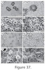
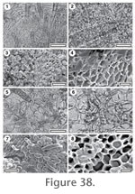
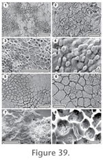
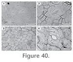
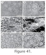
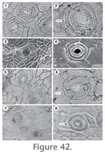
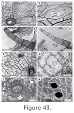
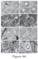
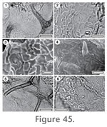
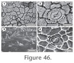
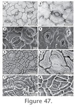
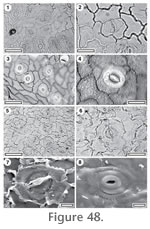
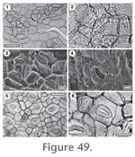
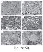
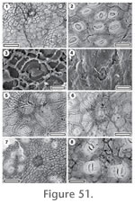
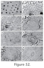
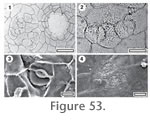
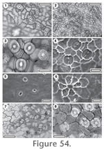
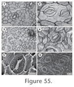
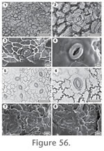 Reference Specimen and locality: SB1379, BL-32.