|
|
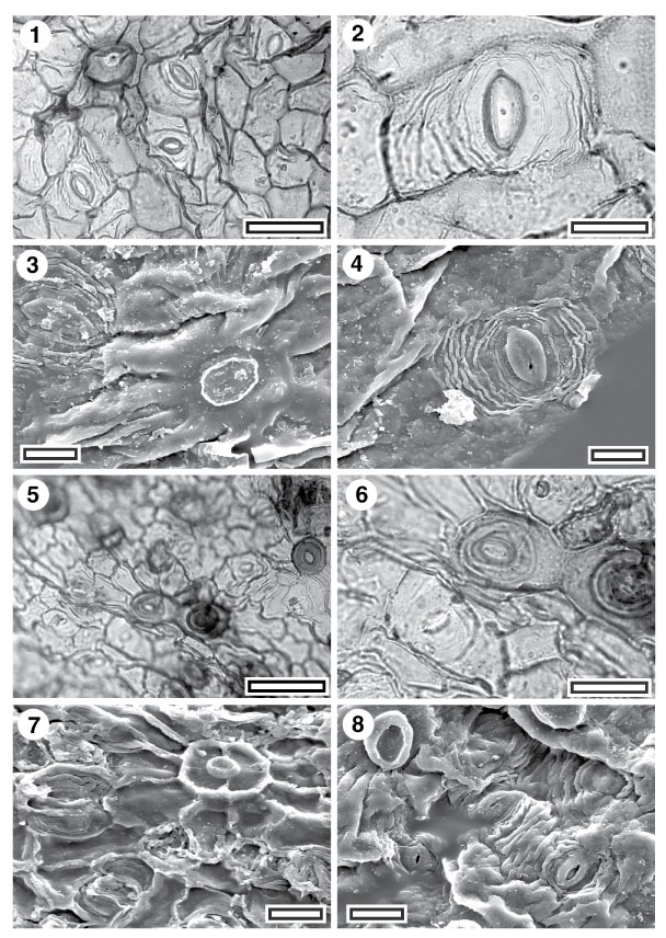
Figure 13. 1-8. Fossil Proteaceae: CUT-P-EHA and CUT-P-EHJ. 1. CUT-P-EHA, TLM view showing stomatal complexes and (upper left) a multi-cellular trichome attachment scar. Note how epidermal cells are partially obscuring parts of some stomatal complexes (SL1680, scale-bar = 50 µm); 2. CUT-P-EHA, TLM detail of single stomatal complex (SL1680, scale-bar = 20 µm); 3. CUT-P-EHA, SEM view of inner cuticular surface showing a single stomatal complex (S-1056, scale-bar = 20 µm); 4. CUT-P-EHA, SEM view of outer cuticular surface showing a single stomatal complex with striae over the subsidiary cells (S-1056, scale-bar = 20 µm); 5. CUT-P-EHJ, TLM view showing stomatal complexes and multi-cellular trichome attachment scars (SL1682, scale-bar = 50 µm); 6. CUT-P-EHJ, TLM detail of single stomatal complex and two multi-cellular trichome attachment scars (SL1682, scale-bar = 20 µm); 7. CUT-P-EHJ, SEM view of inner cuticular surface showing stomatal complexes and (top right) a trichome attachment scar (S-1057, scale-bar = 20 µm); 8. CUT-P-EHJ, SEM view of outer cuticular surface showing stomatal complexes and (upper left) a trichome attachment scar (S-1057, scale-bar = 20 µm).
|