|
|
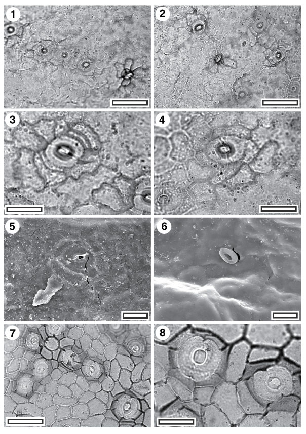
Figure 26. 1-8. Fossil Cunoniaceae-Elaeocarpaceae: CUT-Z-CBI and CUT-Z-FAJ. 1. CUT-Z-CBI, TLM view showing stomatal complexes and (lower right) a trichome attachment scar. Note how outlines of epidermal cells become obscure away from stomatal complexes (SL3049, scale-bar = 50 µm); 2. CUT-Z-CBI, TLM view showing stomatal complexes and (centre) a trichome attachment scar (SL3049, scale-bar = 50 µm); 3. CUT-Z-CBI, TLM detail of single stomatal complex with clear brachyparacytic structure (SL3049, scale-bar = 20 µm); 4. CUT-Z-CBI, TLM detail of single stomatal complex with no apparent brachyparacytic structure (SL3049, scale-bar = 20 µm); 5. CUT-Z-CBI, SEM view of inner cuticular surface showing a single stomatal complex. Note very shallow anticlinal walls of subsidiary cells, and essential absence of walls else where (S-1122, scale-bar = 20 µm); 6. CUT-Z-CBI, SEM view of outer cuticular surface showing a single stomatal complex (S-1122, scale-bar = 20 µm); 7. CUT-Z-FAJ, TLM view showing stomatal complexes. Note giant stomatal complex (lower right) (SL2106, scale-bar = 20 µm); 8. CUT-Z-FAJ, TLM view showing stomatal complexes. The narrow subsidiary cells are well-defined by their thicker periclinal walls (SL2108, scale-bar = 50 µm).
|