|
|
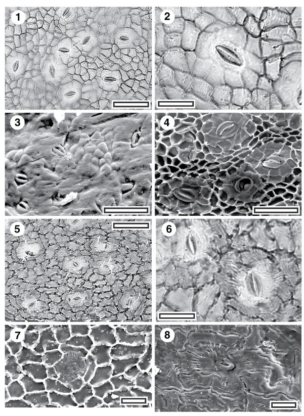
Figure 28. 1-8. Fossil Meliaceae. Note the tendency to have well-defined outer stomatal ledges "floating" within a poorly–outlined stoma: CUT-Z-CDE, and CUT-Z-CDD. 1. CUT-Z-CDE, TLM view showing stomatal complexes (SL0302, scale-bar = 50 µm); 2. CUT-Z-CDE, TLM detail of single stomatal complex (SL0302, scale-bar = 20 µm); 3. CUT-Z-CDE, SEM view of outer surface showing stomatal complexes in a slightly papillate surface (S-1347, AQ 441329, scale-bar = 50 µm); 4. CUT-Z-CDE, SEM view of inner cuticular surface showing four stomatal complexes. Note clearly different texture of subsidiary cells than ordinary epidermal cells, and that by this criteria, not all subsidiary cells are in direct contact with the guard cells (S-1346, B. Hyland 8650, scale-bar = 50 µm); 5. CUT-Z-CDD, TLM view showing stomatal complexes (SL0342, scale-bar = 50 µm); 6. CUT-Z-CDD, TLM view showing two stomatal complexes (SL0342, scale-bar = 20 µm); 7. CUT-Z-CDD, SEM view of inner cuticular surface showing a single stomatal complex (S-1201, scale-bar = 20 µm); 8. CUT-Z-CDD, SEM view of outer cuticular surface showing a single stomatal complex with radiating striae (S-1201, scale-bar = 20 µm).
|