|
|
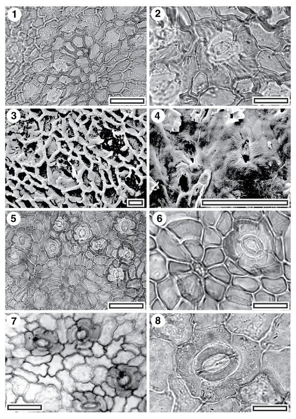
Figure 31. 1-6. Fossil Sapindaceae: CUT-Z-ABF, CUT-Z-CEI and CUT-Z-GDC. 1. CUT-Z-ABF, TLM view showing stomatal complexes and (just to lower right of centre) a trichome attachment scar (SB1382, scale-bar = 50 µm); 2. CUT-Z-ABF, TLM detail of single stomatal complex. Note flanges of subsidiary cells projecting over the outline of the guard cells (SB1382, scale-bar = 20 µm); 3. CUT-Z-ABF, SEM view of inner cuticular surface showing three stomatal complexes (S-1345, NE 043791A, scale-bar = 10 µm); 4. CUT-Z-ABF, SEM view of outer cuticular surface with a clear stomatal complex at upper right. Note short bands of striae radiating from complex (S-1345, NE 043791A, scale-bar = 20 µm); 5. CUT-Z-CEI, TLM view showing stomatal complexes and trichome attachment scars (SL0364, scale-bar = 50 µm); 6. CUT-Z-CEI, TLM detail of single stomatal complex and trichome attachment scar (SL0364, scale-bar = 20 µm); 7. CUT-Z-GDC. TLM view showing stomatal complexes (SL1222, scale-bar = 50 µm); 8. CUT-Z-GDC. TLM detail of single stomatal complex (SL1222, scale-bar = 20 µm).
|