|
|
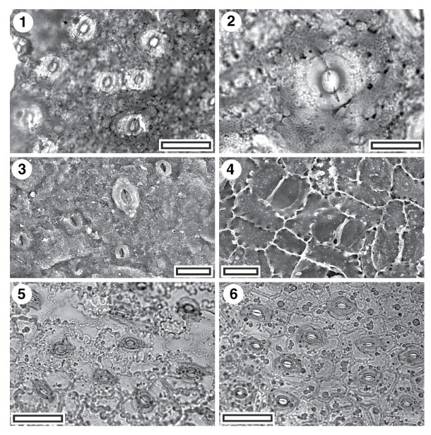
Figure 32. 1-4. Fossil and extant Ericaceae. 1. CUT-Z-ECD, TLM view showing generally stomatal complexes (SL1929, scale aligned -bar = 50 µm); 2. CUT-Z-ECD, TLM detail of single stomatal complex (SL1929, scale-bar = 20 µm); 3. CUT-Z-ECD, SEM view of outer cuticular surface showing (upper centre) a single stomatal complex with prominent outer stomatal ledges, and 4-5 smaller structures which are probably trichome attachment scars (S-1077, scale-bar = 20 µm); 4. CUT-Z-ECD, SEM view of inner cuticular surface showing three stomatal complexes. Note buttressing of walls right up to the margins of the guard cells (S-1077, scale-bar = 20 µm); 5. Extant Trochocarpa laurina, TLM view showing aligned stomata and highly sinuous epidermal cell walls (AQ610328, scale-bar = 50 µm); 6. Extant T. bellendenkerensis, TLM view showing aligned stomata but little sinuosity of epidermal cells (AQ188409, scale-bar = 50 µm).
|