|
|
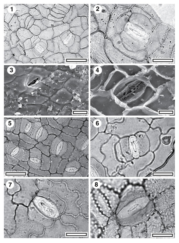
Figure 2. 1-8. Fossil and extant Gnetales. 1. CUT-Z-ADE, TLM view showing stomatal complexes (SL0093, scale-bar = 50 µm); 2. CUT-Z-ADE, TLM detail of single stomatal complex (SL0093, scale-bar = 20 µm). Note the distinctive elongate shape of the guard cell pair, with no sign of separating walls, and no sign of an outer stomatal ledge; 3. CUT-Z-ADE, SEM view of outer cuticular surface showing a single stomatal complex (S-1125, scale-bar = 20 µm); 4. CUT-Z-ADE, SEM view of inner cuticular surface showing a single stomatal complex (S-1125, scale-bar = 10 µm); 5. Extant Gnetum microcarpum, TLM view showing stomatal complexes (AQ142225, scale-bar = 50 µm); 6. Extant G. gnemon, TLM detail of single stomatal complex (AQ142124, scale-bar = 20 µm); 7. Extant G. tenuifolium, TLM detail of single stomatal complex (AQ142229, scale-bar = 20 µm); 8. Extant G. latifolium, TLM detail of single stomatal complex (AQ142207, scale-bar = 20 µm).
|