|
|
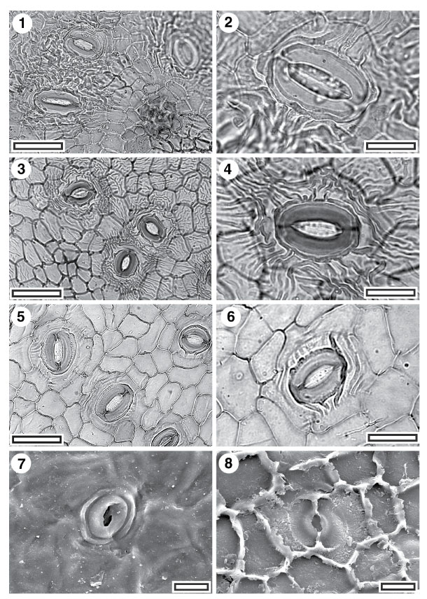
Figure 7. 1-8. Fossil Atherospermataceae: CUT-Z-CFA. 1. TLM view showing stomatal complexes and a massively-thickened trichome attachment scar (SB1373, scale-bar = 50 µm); 2. TLM detail of single stomatal complex (SB1373, scale-bar = 20 µm). Note the distinctive outer stomatal rim; 3. TLM view showing stomatal complexes (SL2960, scale-bar = 50 µm); 4. TLM detail of single stomatal complex (SL2960, scale-bar = 20 µm). Note the distinctive outer stomatal rim; 5. TLM view showing stomatal complexes (SL0304, scale-bar = 50 µm); 6. TLM detail of single stomatal complex (SL0304, scale-bar = 20 µm). Note the distinctive outer stomatal rim; 7. SEM view of outer cuticular surface showing a single stomatal complex with prominent outer stomatal ledges surrounded by a discontinuous rim (S-1096, scale-bar = 20 µm); 8. SEM view of inner cuticular surface showing a single stomatal complex. Note granular texture of subsidiary cell periclinal walls compared with epidermal cells (S-1096, scale-bar = 20 µm).
|