|
|
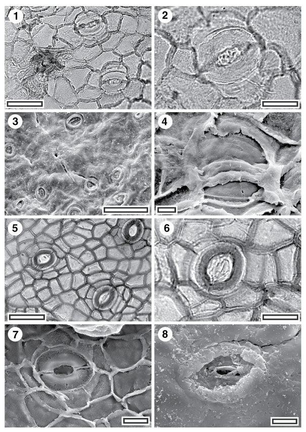
Figure 9. 1-8. Fossil Atherospermataceae: CUT-Z-JIF and CUT-Z-ECG. 1. CUT-Z-JIF, TLM view showing stomatal complexes and a massively-thickened trichome attachment scar (SB0851, scale-bar = 50 µm); 2. CUT-Z-JIF, TLM detail of single stomatal complex (SB0851, scale-bar = 20 µm); 3. CUT-Z-JIF, SEM view of outer cuticular surface showing stomatal complexes and (just left of centre) a trichome attachment scar (S-319, scale-bar = 0.1 mm); 4. CUT-Z-JIF, SEM view of inner cuticular surface showing a single stomatal complex (S-319, scale-bar = 10 µm); 5. CUT-Z-ECG, TLM view showing stomatal complexes (SL1954, scale-bar = 50 µm); 6. CUT-Z-ECG, TLM detail of single stomatal complex (SL1954, scale-bar = 20 µm); 7. CUT-Z-ECG, SEM view of inner cuticular surface showing a single stomatal complex (S-1073, scale-bar = 20 µm); 8. CUT-Z-ECG, SEM view of outer cuticular surface showing a single stomatal complex (S-1073, scale-bar = 10 µm).
|