| |
INTRODUCTION
Optical microscopy remains the preferred method for imaging in paleopalynology. The study of palynomorphs, for both taxonomic description and quantitative data extraction from counts, strongly relies on the observation of palynological assemblages mounted on microscope slides using traditional techniques of preparation (Doher 1980;
Wood et al. 1996;
Traverse 2007). With the improvement of optical microscopy over time, imaging of details on transparent samples (which until then had often produced poor quality results) was made possible by optical means, without the use of chemical/dying techniques (Pluta 1989;
Slayter and Slayter 1992;
Davidson and Abramowitz 2002), which alter the subject in some way. These innovations include various contrast enhancing techniques (Hoffman 1977;
Abramowitz 1987;
Bradbury and Evennett 1996;
Davidson and Abramowitz 2002) and more especially differential interference contrast (DIC) microscopy, invented in the mid 1950s (Allen et al. 1969;
Nomarski 1955). For palynology, DIC allows the observation of very minute ornamentation of the exine not visible under regular brightfield microscopy.
 However, optical microscopy suffers from resolution limitations. Horizontal resolution limitation (the resolving power between two points occurring in the plane perpendicular to the optical axis) is often given and is an easily understood parameter as it directly translates into maximum attainable magnification of an object. Additionally, optical systems are also characterized by their ability to resolve along the optical axis, which is termed axial resolution. The second resolution, measured in a plane parallel to the optical axis, is known as the depth of field (Pluta 1989;
Slayter and Slayter 1992;
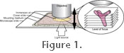
Davidson and Abramowitz 2002). Practically, depth of field represents the distance that separates the nearest object plane in focus to the farthest object plane which is simultaneously in focus (Davidson and Abramowitz 2002). In microscopy, this distance is very short and typically measured in microns. This very limited depth of field does not present an issue for flat objects such as thin section, but objects preserved in three dimensions (such as palynomorphs), with a greater thickness than the depth of field for the objective. In such cases it is only possible to see a single optical cross section of the object at a time, and the entirety of the object can only be reconstructed as a mental image by constantly varying the focus adjustment of the microscope (Figure 1). However, optical microscopy suffers from resolution limitations. Horizontal resolution limitation (the resolving power between two points occurring in the plane perpendicular to the optical axis) is often given and is an easily understood parameter as it directly translates into maximum attainable magnification of an object. Additionally, optical systems are also characterized by their ability to resolve along the optical axis, which is termed axial resolution. The second resolution, measured in a plane parallel to the optical axis, is known as the depth of field (Pluta 1989;
Slayter and Slayter 1992;
Davidson and Abramowitz 2002). Practically, depth of field represents the distance that separates the nearest object plane in focus to the farthest object plane which is simultaneously in focus (Davidson and Abramowitz 2002). In microscopy, this distance is very short and typically measured in microns. This very limited depth of field does not present an issue for flat objects such as thin section, but objects preserved in three dimensions (such as palynomorphs), with a greater thickness than the depth of field for the objective. In such cases it is only possible to see a single optical cross section of the object at a time, and the entirety of the object can only be reconstructed as a mental image by constantly varying the focus adjustment of the microscope (Figure 1).
Both axial and horizontal resolutions are driven by the numerical aperture of the objective, but in different ways. As the axial resolution decreases, the horizontal resolution increases with the numerical aperture. Therefore, objectives with higher numerical apertures give more contrast and higher magnification but a lower depth of field, thus it is necessary to select an appropriate trade-off between these quantities. Traditionally this trade-off is solved in photographic descriptions of palynomorphs by including a general view of the specimen taken at a lower magnification to avoid depth of field problems, and a series of pictures at higher magnification to highlight details, or to expose different views of the same specimen as separate optical sections. While the inclusion of multiple pictures for a single palynomorph is necessary and commonly used for taxonomically oriented publications (such as for description of new species), it is rarely done in publications involving description of palynological assemblages for biostratigraphy.The amount of space required for depicting detailed palynological plates can be quite high for large assemblages, and multiple views of a single palynomorph for identification is not practical. As a result, palynomorphs are depicted as a single lower resolution photograph that may not show all the necessary details for easy identification and verification.
To circumvent this problem, dedicated software solutions may be employed to reconstruct a single image from multiple optical sections, each containing only parts of the object in focus. This technique allows obtaining the advantage of higher magnification and contrast levels given by the highest magnification objectives, whilst eliminating the limited depth of field issue.
|