![]()
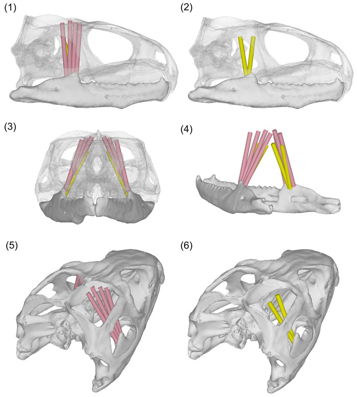
FIGURE 5. mPstS (pink) and mPstP (yellow). (1) Lateral view showing both the mPstS and mPstP with the skull at 80% transparency; (2) lateral view showing only the mPstP with the skull at 80% transparency; (3) anterior view showing both the mPstS and mPstP with the skull at 80% transparency; (4) posterolateral view of only the lower jaw showing both the mPstS and mPstP; (5) posterodorsolateral view of the mPstS; (6) posterodorsolateral view of the mPstP. The fascia is not included in this figure.