![]()
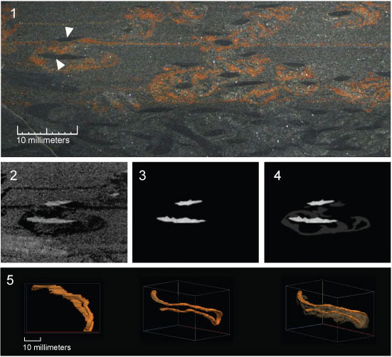
FIGURE 6. Image processing stages during three-dimensional reconstruction of phycosiphoniform from Rosario Formation. 6.1. Investigated material from Rosario Formation containing phycosiphoniform forms. Each photograph obtained during serial grinding (set of 60 sequential images) was aligned and cropped. The continuous burrow cores were selected manually as the object of study (indicated by white arrows). 6.2. To improve contrast, images were converted to gray scale. 6.3. Distinct burrow cores were manually selected using layer masks in Photoshop and hiding all other burrow cores in the investigated area of the original images. Selected cores were tracked on all processed images. Images were then cropped to size that encompassed isolated burrow cores on all processed images. 6.4. For additional reconstruction of burrow with its surrounding halos, areas of the halos were manually marked on all sequential images uncovering it from the masked layer. 6.5. Three-dimensional visualisation of phycosiphoniform obtained through volume rendering of sequential images imported to VolView software.