![]()
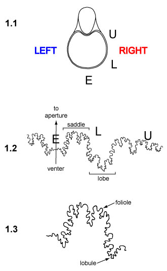
FIGURE 1. Illustration of a typical ammonite suture, with anatomical terms and orientations. 1.1 View of ammonoid shell, facing the aperture. Sutures are divided into external (E), lateral (L), umbilical (U), and internal (not shown) elements, moving from a ventral to lateral to dorsal position. Diagram of shell is redrawn and modified from Monks and Palmer (2002, p. 44). 1.2 Ammonite suture pattern with key elements labeled. The venter or outer central margin of the shell is indicated by an arrow; the arrow points toward the aperture and soft body of the animal. Note that, given this traditional orientation, the "right" suture (i.e., drawn to the right of the arrow; labeled "RIGHT" in 1.1) actually represents the left side of the ammonoids body. Portions of the suture line that fold up towards the aperture are called saddles; portions that fold down toward the shell apex are called lobes. Suture pattern is from Neogastroplites muelleri, USNM 129456, digitized from Reeside and Cobban (1960), figure 19f. 1.3 Close-up view of portion of lateral element figured in 1.2. Subfolds on saddles are termed folioles, while subfolds on lobes are termed lobules.