![]()
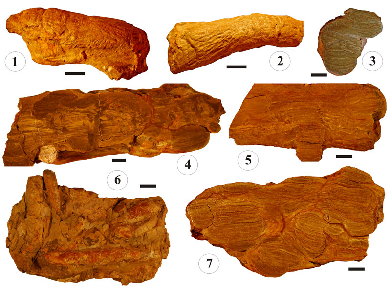
FIGURE 10. Close-ups of Trichophycus venosus. 1. Specimen with coarse longitudinal scratch marks clearly visible in the lower part of the structure, and finer, oblique scratch marks on the sides of the structure. 2. Same specimen as in 1, showing coarse longitudinal scratch marks and transversal constriction-like ridges locally develop (right). 3. Detail of spreite structure. Sand laminae are separated by thin stylolytes. 4. Tempestite displaying microhumocky cross-stratification. Note the presence of escape structures (center left). The cluster structure on the upper left is Phycodes isp. (note the spreite laminae are mud-dominated rather than sand-dominated). Trichophycus specimen on the left also displays mud-rich spreite laminae. 5. Trichophycus displaying a distinctive flat spreite (center). The other two specimens, to the right and to the left, are camouflaged with the host rock (i.e., similar lithology). 6. Base showing high density of Trichophycus surrounded by fine-grained siltstone. 7. Cross-sectional view showing a thoroughly bioturbated silty sandstone. Note that almost a 100% of the polished surface corresponds to Trichophycus spreite. Scale bars equal 1 cm.