![]()
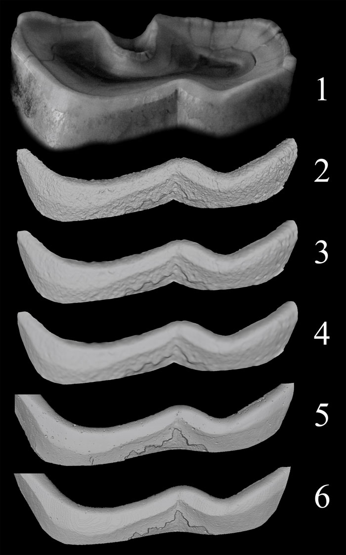
FIGURE 2. Comparison of the models corresponding to the buccal side enamel edge of D. bicornis FMNH-945/1960. 1. photograph of the upper part of m2d; 2. photograph-based 3D model before smoothing; 3. photograph-based 3D model after 3 steps of smoothing; 4. photograph-based 3D model after 7 steps of smoothing and after vertex removal; 5. 3D model from laser scanner (30 μm nominal resolution); 6. 3D model from needle scanner (50 μm nominal resolution).