![]()
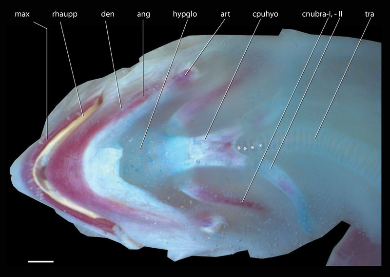
FIGURE 19. Emydura subglobosa. 1) Ventral view of the head of a cleared and double stained subadult specimen (PIMUZ lab# 2009.9). Red = calcified tissue, blue = cartilaginous tissue, light blue = connective tissue, yellow = non stained keratin. Note the different degree of ossification of the bones of the hyoid apparatus; especially the cornu branchial-II has a very low amount of ossification. Asterisks indicate ossification centres in the tracheal rings.