![]()
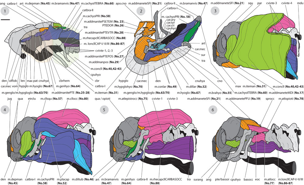
FIGURE 23. Continuation of Figure 22. 1) ventral view to the lower jaw and hyoid region, mm. intermandibularis (No. 31) and constrictor colli (No. 40, 42-43) are transparent, 2) dorsolateral and frontal view to the lower jaw and the hyoid region, 3-6) different layers of neck and hyoid muscles, 3) all neck muscles are shown, m. constrictor colli (No. 40, 42-43) is partly removed in in the posterodorsal region to enable a view to the dorsal aspects of the cervical vertebrae, 4) m. constrictor colli (No. 40, 42-43) and m. intermandibularis (No. 31) removed, 5) further neck muscles removed, 6) most neck muscles removed highlighting the origin sites of m. adductor mandibulae externus (No. 17, 19, 21). Bar scale (under "1"): for 1, 3-6) ca. 0.5 mm; for 2) ca. 0.25 mm.