![]()
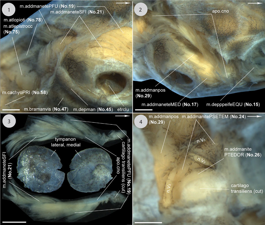
FIGURE 29. Continuation of Figure 28; continued in Figure 30, Figure 31. 1) Dorsolateral view to the posterior region of the head showing the origin sites of m. adductor mandibulae externus (No. 19, 21). Removed: mm. collooccipitis (No. 80) et plastrocapitis (No. 52), et collosquamosus (No. 57). 2) Dorsal view to the adductor chamber demonstrating the course of the coronar aponeurosis, and the orientation of mm. adductor mandibulae externus Pars medialis (No. 17) et adductor mandibulae posterior (No. 29). Removed: jugal and postorbital, m. collooccipitis (No. 80), the bone bridge made of parts of squamosal and parietal, 3) The coronar aponeurosis in lateral (top) and medial (bottom) view with most fibres of m. adductor mandibulae externus (No. 19, 21) removed, cut within cartilago transiliens. 4) Dorsolateral view into the postorbital cavity of the adductor chamber. Removed: m. adductor mandibulae externus (No. 17, 19, 21) - cut within cartilago transiliens, postorbital, jugal. Scale equals 2 mm.