![]()
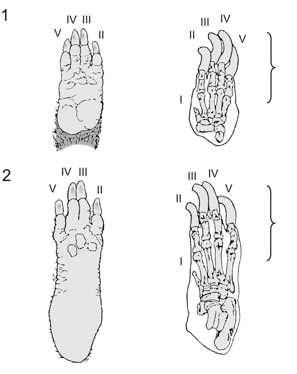
FIGURE 7. Foot structure of Dinomys branicki. 1. Forefoot structure, to the left on plantar view, to the right on dorsal view. 2. Hind foot structure, to the left on plantar view, to the right on dorsal view. Note the printed area of the foot (the digits and the metapodial-phalangeal junctions) on the substrate. Modified from Grand and Eisenberg (1982).