![]()
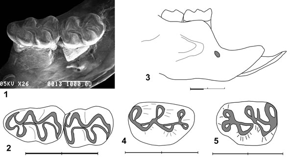
FIGURE 43. 1. Peromyscus sp. A (UCMP 320570), occlusal view of M1-M2 in right dentary fragment. SEM image. Scale equals 1 mm. 2. Peromyscus sp. A (UCMP 320570), occlusal view of M1-M2. SEM image. Scale equals 2 mm. 3. Peromyscus sp. A (UCMP 320570), labial view of right dentary. SEM image. Scale equals 2 mm. 4. Peromyscus sp. A (UCMP 320574), occlusal view of M1. SEM image. Scale equals 2 mm. 5. Peromyscus sp. B (UCMP 320572), occlusal view of M1. SEM image. Scale equals 2 mm.