FIGURE 1. Geological map of the continental sediments of the Tudela Formation in the Bardenas Reales de Navarra area, with location of the Pico del Fraile section and the Pico del Fraile 1 (PF1) locality. The location of other fossil localities studied previously by Murelaga (2000) and Murelaga et al. (2004a and 2004b) is also shown.
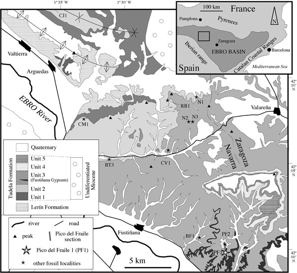
FIGURE 2. Composite lithostratigraphic and magnetostratigraphic logs of the Tudela Formation (see Larrasoaña et al., 2006) and their correlation to the ATNTS2004 of Lourens et al. (2004). The position of locality PF1 is described in this study; for the other fossil localities see Murelaga (2000) and Murelaga et al. (2004a, 2004b).
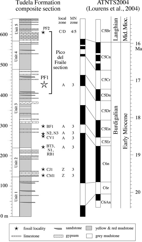
FIGURE 3. Nomenclature of parts of the cheek teeth, adapted after Freudenthal (2004).
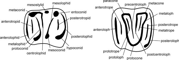
FIGURE 4. Vasseuromys rambliensis sp. nov. from PF1, Ebro basin, Spain. 1, PF1 29, left d4; 2, PF1 30, left d4; 3, PF1 28, left p4; 4, PF1 15 (holotype), right m1; 5, PF1 8, right m2; 6, PF1 12, left m2; 7, PF1 16, right m2; 8, PF1 9, right m3; 9, PF1 10, right m3; 10, PF1 11, right m3; 11, PF1 24, left D4; 12, PF1 27, right D4; 13, PF1 37, right P4; 14, PF1 2, left M1; 15, PF1 1, right M2; 16, PF1 3, left M2; 17, PF1 58, left M2; 18, PF1 36, left M3. Scale bar equals 1 mm.
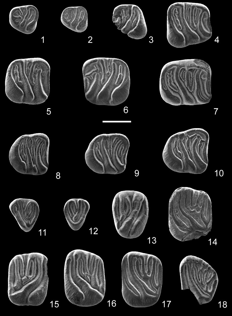
FIGURE 5. Ranges of variation of length and width (tenths of millimeters) of the lower molars of V. rambliensis sp. nov. from PF1, V. pannonicus from Eichkogel, V. elegans from Stubersheim 3, V. duplex from Harami 1, V. bacchius from Fuenmayor 2, V. rugosus from Santa Cilia and Laugnac, V. priscus from Santa Cilia and V. autolensis from Autol 1.
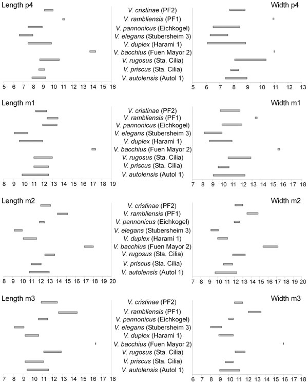
FIGURE 6. Ranges of variation of length and width (tenths of millimeters) of the upper molars of V. rambliensis sp. nov. from PF1, V. pannonicus from Eichkogel, V. elegans from Stubersheim 3, V. duplex from Harami 1, V. bacchius from Fuenmayor 2, V. rugosus from Santa Cilia and Laugnac, V. priscus from Santa Cilia and Moissac 1 and V. autolensis from Autol 1.
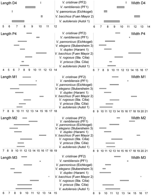
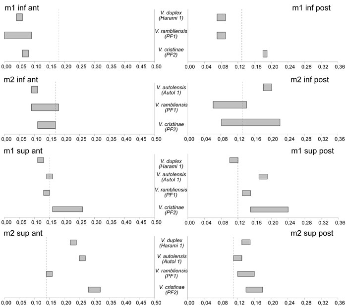
FIGURE 7. Ranges of concavity of anterior and posterior side of m1, m2, M1 and M2 of Vasseuromys duplex from Harami 1, V. autolensis from Autol 1, V. rambliensis sp. nov. from PF1 and V. cristinae from PF2. The dashed lines indicate the limits between weakly and moderately concave (from Freudenthal and Martín-Suárez, 2007b).