FIGURE 1.Summary table of the geochronological positioning of the Teresina Formation (subdivision proposed by Walker and Geissman, 2009).
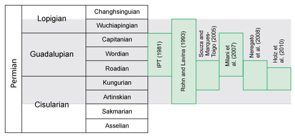
FIGURE 2. Geological and location map of the study area. Geological base: CPRM (2006).
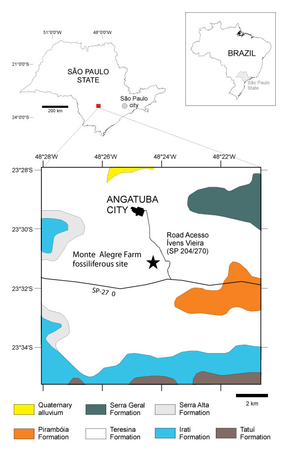
FIGURE 3. 3.1 - General view of the location where silicified stromatolite blocks occur; 3.2 - Blocks of silicified stromatolites; 3.3 - Outcrops in the Acesso Ivens Vieira-Angatuba road cutting, with large silicified bioherms at the base (Figure 3.4) and silicified levels (S).
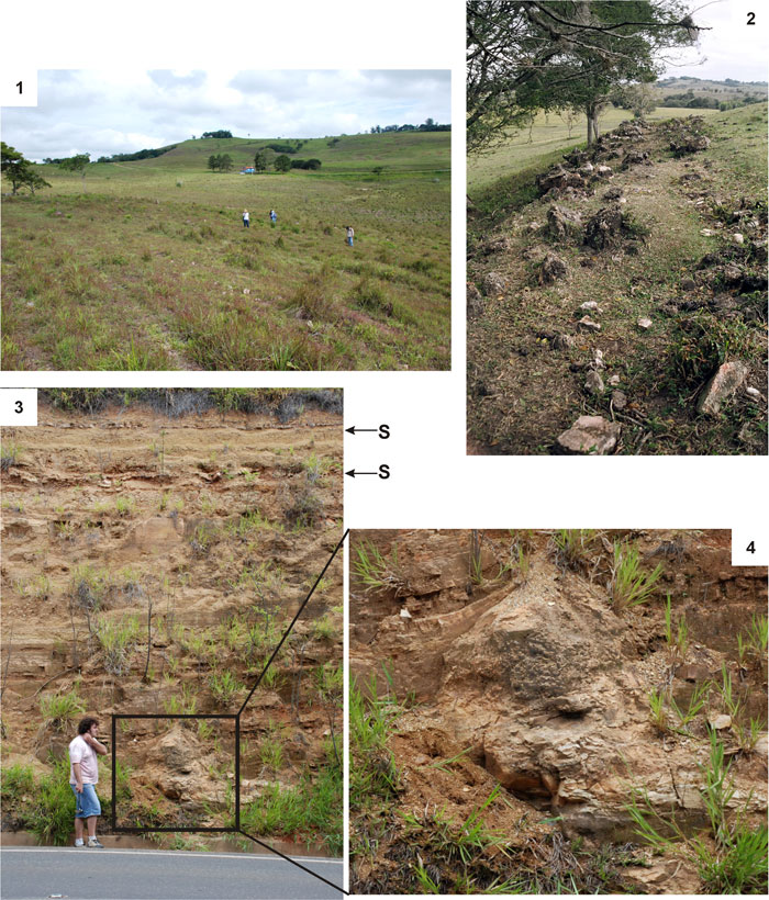
FIGURE 4. 4.1 - Domed bioherm; 4.2 and 4.3 - Plan view of the lenticular bioherm.
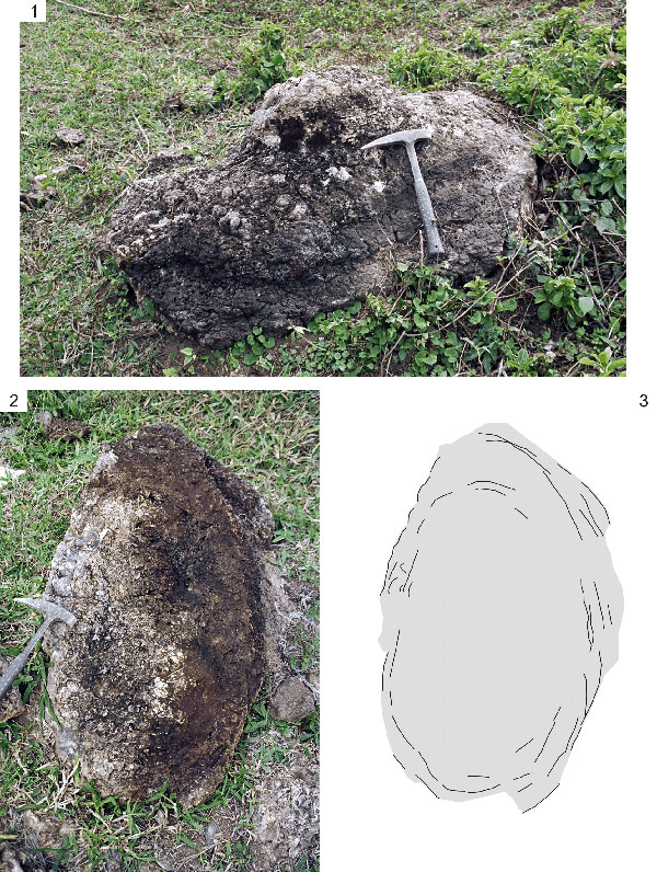
FIGURE 5. Bioherm with structure in the form of "scales" at the top (5.1) and macrostructure of anastomosed, non-isolated multiple columns showing parallel to divergent growth and a laminar profile ranging from slightly convex to parabolic in the longitudinal section (5.2-3); 5.4 - Three-dimensional model of the bioherm (Drawing by Raphael Galassi de Amorim).
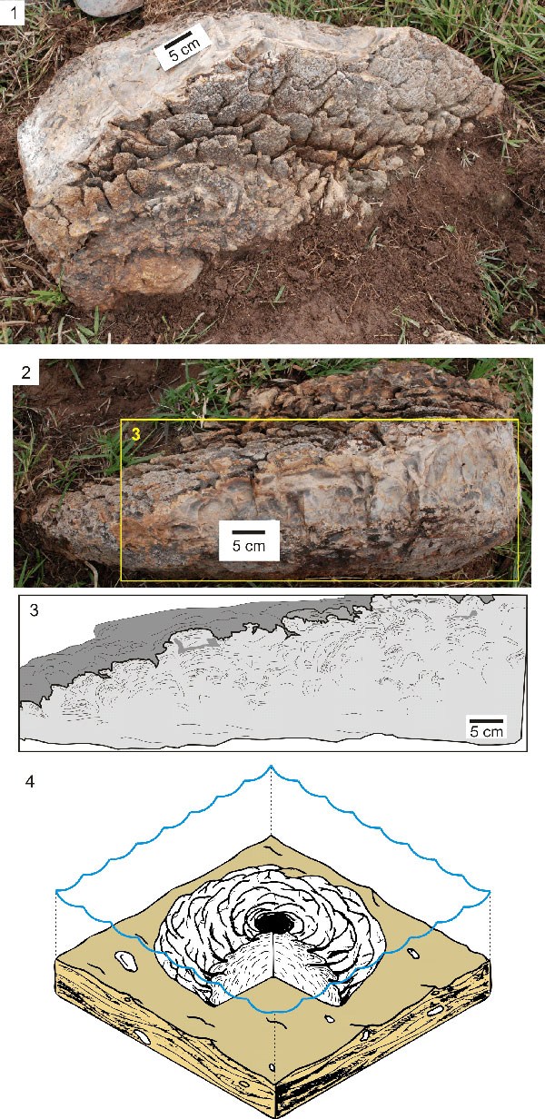
FIGURE 6. 6.1 - Concentric structures at the top of the bioherm; 6.2 - Botryoidal texture at the top of the bioherm.
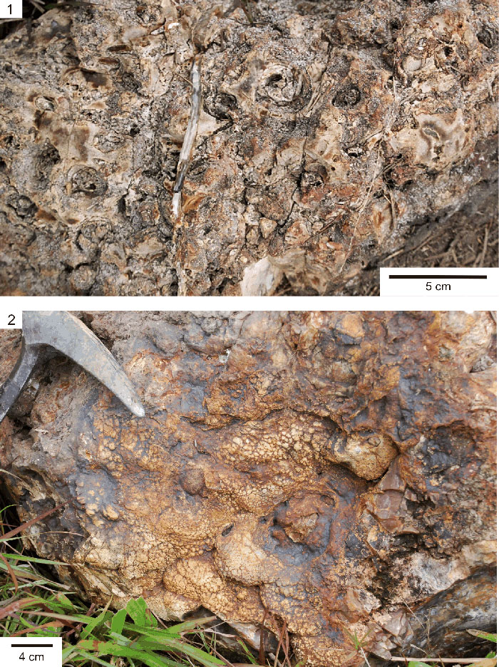
FIGURE 7. 7.1 and 7.2 - Individual columns with convex to parabolic lamination; 7.3 and 7.4 - Weathered bioherm exhibiting multiple columns and fine stromatolitic lamination with alternating light and dark laminas on a petrographic slide (7.4).
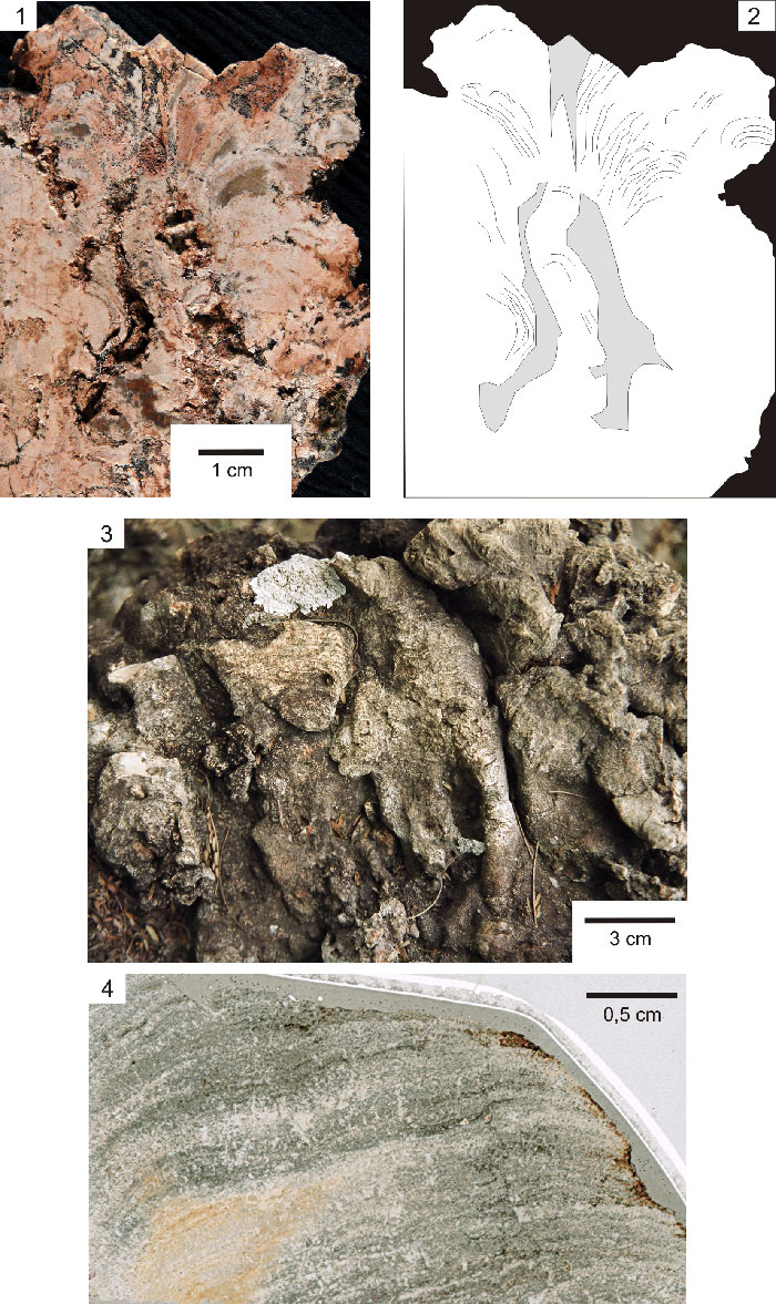
FIGURE 8. 8.1 - Microcolumns forming a botryoidal texture at the top of a bioherm; 8.2 - Longitudinal section showing lamination and a divergent arrangement of microcolumns; 8.3 - Fine stromatolitic lamination on a petrographic slide; 8.4 - Longitudinal petrographic slide showing the lamination and arrangement of microcolumns; 8.5 and 8.6 - Filamentous cyanobacteria arranged perpendicular to the lamination; 8.7 - Microcoleus; 8.8 - Rivularia; 8.9 - Detail of Rivularia showing a preserved mucilaginous sheath and cell wall.
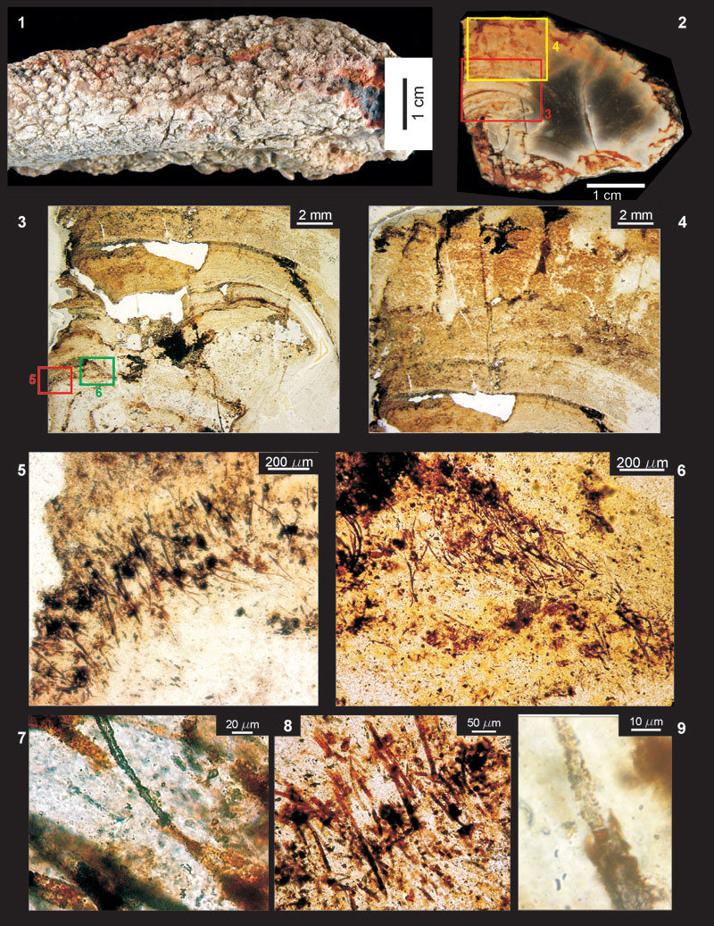
FIGURE 9. SEM images. 9.1 - Fossil of filamentous bacterium (secondary electrons); 9.2 - Fossil of 9.1 mapped by silica content via EDS (red) showing the high contents of the matrix and cell wall (backscattered electrons); 9.3 - Detail of a silicified cell wall and partially hollow interior (secondary electrons).
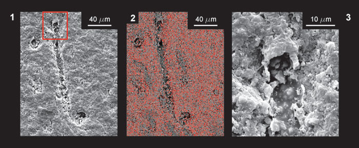
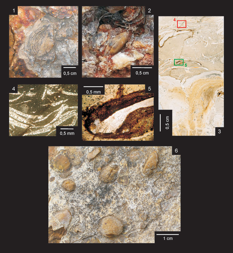
FIGURE 10. 10.1 - Angatubia cowperesioides; 10.2 - Articulated shell of Pinzonella illusa isolated at the top of a bioherm; 10.3 - Detail of coquina and top of the bioherm on a petrographic slide; 10.4 - Detail of coquina on a slide (10.3) with crossed nicols; 10.5 - Detail of stromatolitic overgrowth in bioclasts on a slide of coquina (10.3); F - Upper view of the fossiliferous concentration.