FIGURE 1. A reconstruction of Camptophyllia eltringhami from Gill (1924).
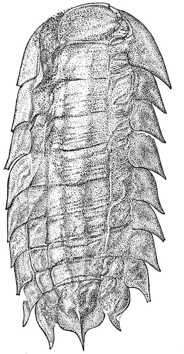
FIGURE 2. Fossil examples of the genus Camptophyllia. 1, NHM In41505, a specimen from the Tyne Coalfield. Visible fossil 44 mm in length. 2, NHM In 41503, the holotype of Camptophyllia fallax Gill 1924, Tyne. Fossil 28 mm in length. 3, Counterpart to 1 showing only posterior segments. Fossil 30 mm in length. 4, NHM In 41504, the holotype of Camptophyllia eltringhami Gill 1924, Tyne. Fossil 39 mm in length. 5, CH3, a specimen from the private collection of Mr. Stephen Livesley, Crock Hey. Fossil 42 mm in length. 6, CH304a, a specimen from the private collection of Mr. Sean Sale, Crock Hey. Fossil 35 mm in length. 7, NHM I. 13951, a specimen from Coseley. Fossil 18 mm in length. 8, NHM I. 13952, a specimen from Coseley. Fossil 13 mm in length. 9, NHM In 22843, a specimen from Coseley. Fossil 20 mm in length. 10, NHM In 22844, a specimen from Coseley. Visible fossil 20 mm in length.
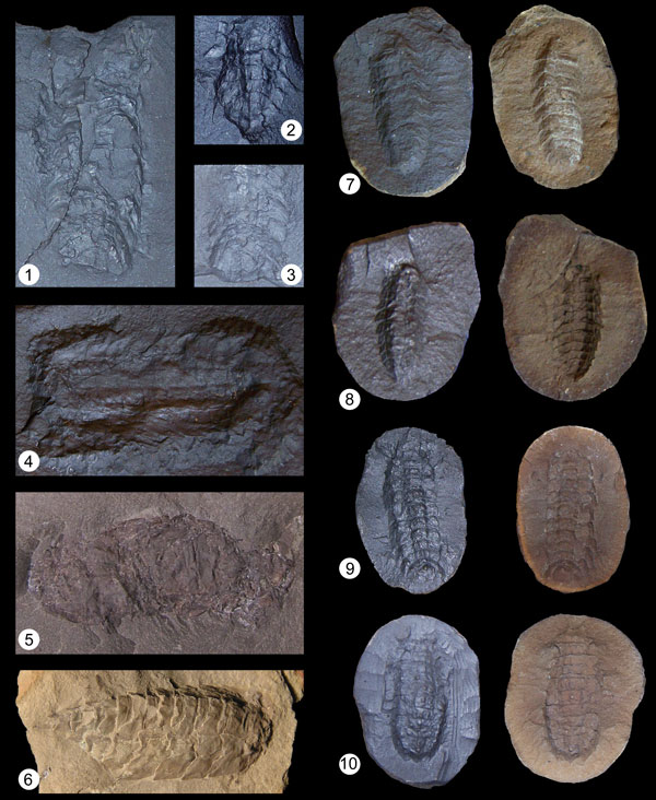
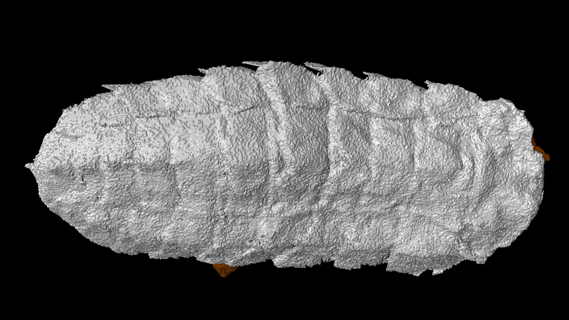
FIGURE 3. Computer reconstruction of Camptophyllia specimen NHM In 22843 from Coseley. 1, Dorsal view. 2, Lateral view. 3, Ventral view. 1-10 = segment numbers; AR = artefact; LS = lateral spine; LT = lateral tubercle; TR = transverse (cephalic) ridge; TT = terminal tubercle. Fossil 20 mm in length.
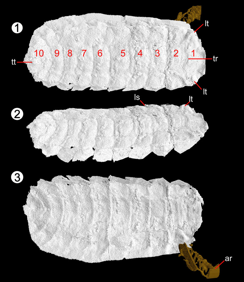
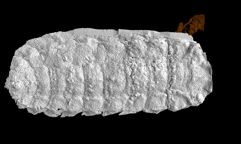
FIGURE 4. Computer reconstruction of Camptophyllia specimen NHM I. 13952 from Coseley. 1, Dorsal view; note lateral curvature of specimen visible from this orientation. 2, Lateral view showing dorsal curvature.Posterior margin of overlapping lateral plates marked by red dashed lines. 3, Zoom of dorsal surface of segments 5-7 showing more complex tergal boundaries accommodating dorsal curvature. 4, Ventral view. 1-10 = segment numbers; ABR = anterior boundary ridge; AR = artefact; DC = line showing zone of dorsal curvature (coiling); LM = lateral margin (ventral); LP = lateral plate; LS = lateral spine; LT = lateral tubercle; M = median depression; MM = median margin (ventral); MS = median spike; MT = median tubercle; PBR = posterior boundary ridge; TR = transverse (cephalic) ridge; TS = terminal spike; TT = terminal tubercle. Fossil is 12.9 mm in length.
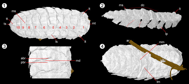
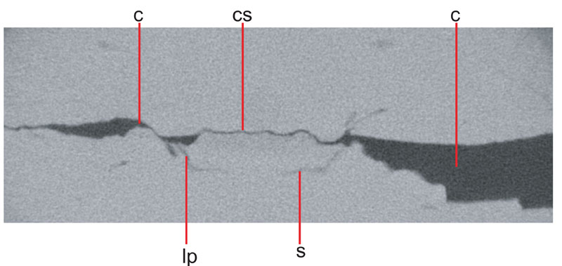
FIGURE 5. XMT slice image through cephalic region of NHM In 22843, showing box-like arrangement of head. C = crack; CS = central sclerite; LP = lateral plate; S = sternite; T = tergite. Fossil is 3.46 mm across.
Animation 1. Tomographic reconstruction of Camptophyllia specimen NHM I.13952 from Coseley.
Animation 2. Tomographic reconstruction of Camptophyllia specimen NHM In 22843 from Coseley.