FIGURE 1. Flow chart showing the fundamental steps in the preparation of VPS replicas for SEM examination and the trapezium-shaped pieces of VPS mounted on geological microscope slides.
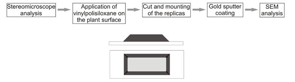
FIGURE 2. 1. VPS replica of the lycopsid Mesenteriophyllum kotschnevii showing isodiametric to transversely elongated epidermal cells. Scale bar equals 100 μm. 2. VPS replica of the bryophyte Muscites brickiae showing longitudinally elongated epidermal cells. Scale bar equals 50 μm. 3. Detail of elongated epidermal cells in the sphenopsid Equisetites obtained with VPS. Scale bar equals 50 μm. 4. Latex replica of the pteridosperm Ptilozamites showing a fissure on the surface. Scale bar equals 200 μm. 5. VPS replica of Ptilozamites showing longitudinal striations on the rachis. Scale bar equals 100 μm. 6–7. Details of stomata in Ptilozamites showing ring-like structure of the subsidiary cells. Scale bars equals 5 μm.
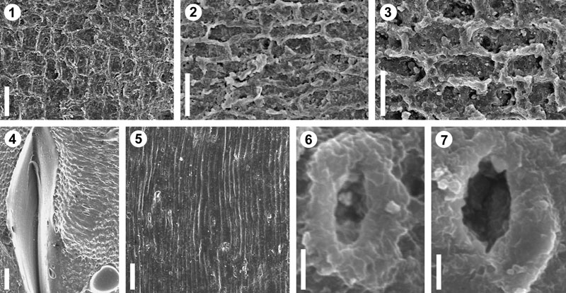
FIGURE 3. 1. SEM image of latex replica of the herbaceous lycopsid Isoetites. Scale bar equals 250 μm. 2. SEM image of VPS replica of the same specimen at the same magnification as shown in Figure 2.1., showing excellent well-preserved epidermal cells. Scale bar equals 250 μm. 3. Detail of the Figure 2.1. Note that details of epidermal cells are indistinguishable, and air bubbles are present over the surface. Scale bar equals 50 μm. 4. Detail of the Figure 2.2. Scale bar equals 50 μm. 5. SEM image of latex replica of the bennettitalean Pterophyllum pinnatifidum. Deformation on the latex surface and air bubbles are present. Scale bar equals 100 μm. 6. SEM image of VPS replica of the same specimen at the same magnification as shown in Figure 2.5., showing intercostal field densely covered with papillae. Scale bar equals 100 μm. 7. Detail of Figure 2.5. Scale bar equals 25 μm. 8. Detail of Figure 2.6., showing a papillate surface. Scale bar equals 25 μm.
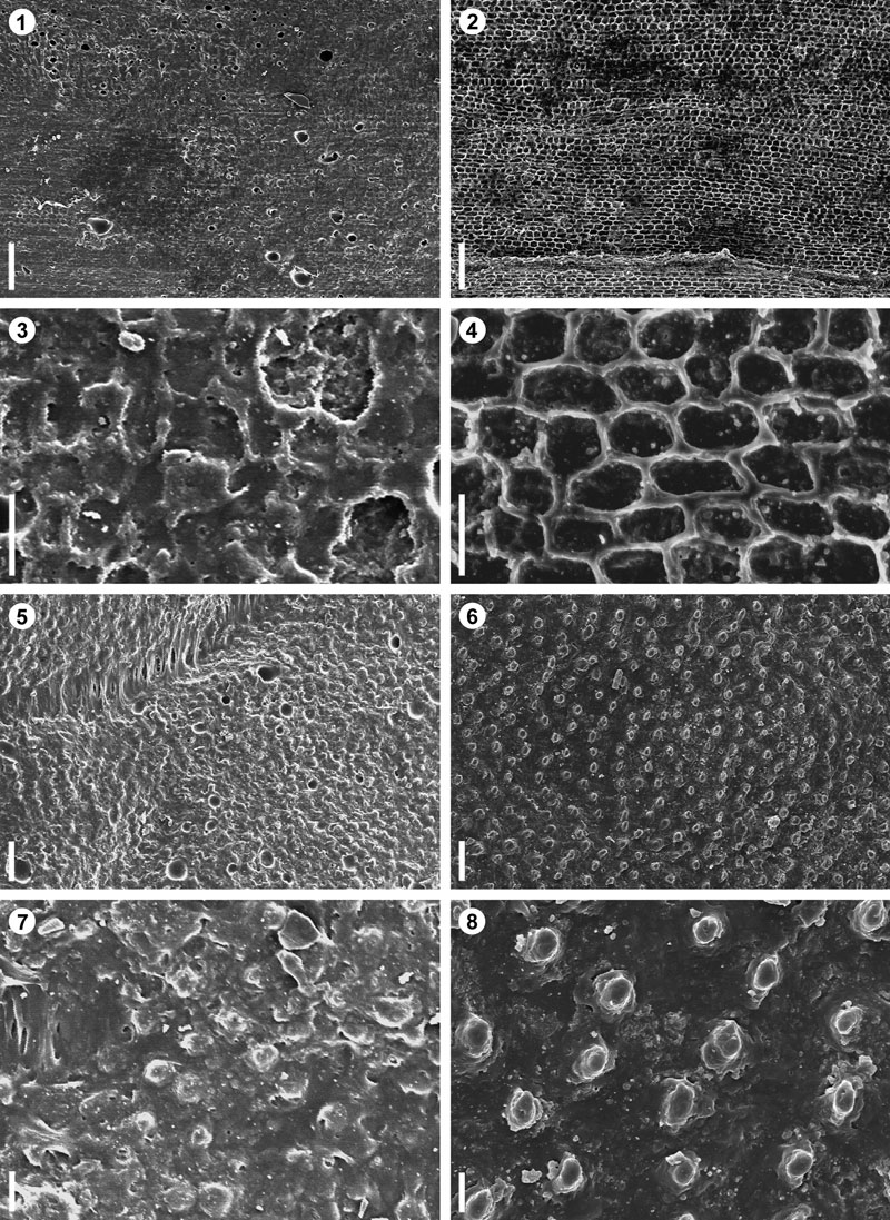
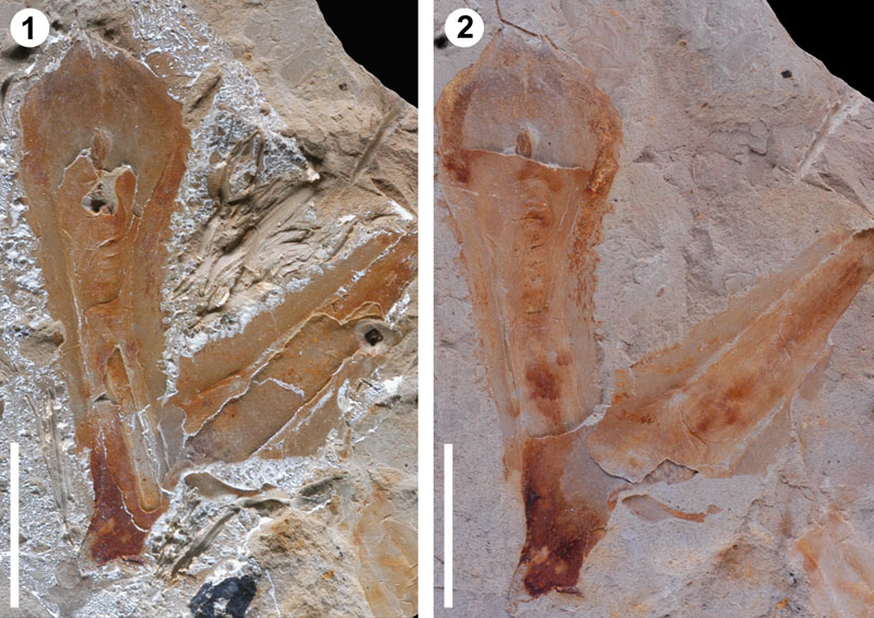
FIGURE 4. Sporophylls of Lepacyclotes zeilleri after being treated with latex (left) and after silicone (right) for SEM examination. Scale bar equals 1 cm.