FIGURE 1. Comparison of gladius shapes between older (1.1, 1.3) and younger stages (1.2, 1.4) of the same species of Coleoidea. 1.1-2. Gladii of Onykia robusta (Verrill, 1876). Gladius of younger stage much stouter than that of older stage. Redrawn after Tsuchiya and Okutani (1991, figures 18, 19). 1.3-4. Gladii of Pholidoteuthis massyae (Pfeffer, 1912), exhibiting shape differences between the different stages. Redrawn after O'Shea et al. (2007, figures 36, 37).
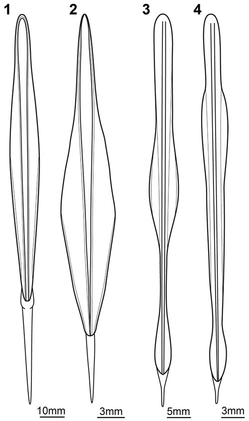
FIGURE 2. The supposed gladius of a fossil cephalopod paralarva (SMNS 67904b), documented with different methods. 2.1. Macrograph under North-West lighting. 2.2. Autofluorescence micrograph. Note the lateral field and the pits, forming a reticulate pattern. 2.3. Red-cyan stereo image with pronounced keel and pits; use red-cyan glasses to view. Abbreviations: ap = apex; ke = keel; lf = lateral field; p = pitted area.
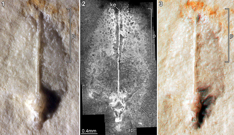
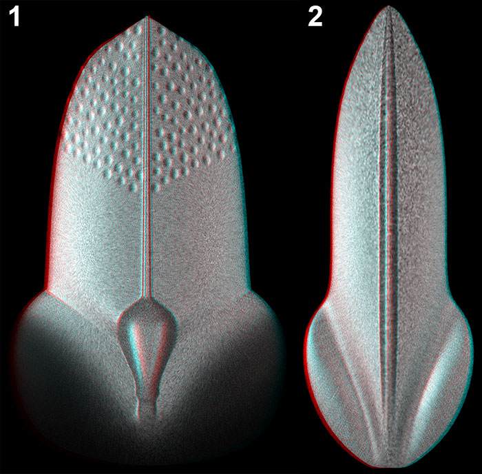
FIGURE 3. 3D models of the gladii of the supposed new paralarva and its posssible adult (red-cyan stereo images). Images not to scale to show the different length-width ratios. 3.1. Gladius of the possible paralarva presented in this paper. The faint areas represent the only partially preserved lateral fields. 3.2. Gladius of the keeled type of Trachyteuthis with morphological similarities to that of the possible paralarva.