FIGURE 1. Location map showing the distribution of the occurrence of the materials mentioned in this contribution. 1. Detail of Patagonia. 2. Detail of Antarctic Peninsula. Abbreviations: 1: Neuquensaurus australis, N. robustus, Laplatasaurus araukanicus, Microcoelus patagonicus, "Titanosaurus" nanus; 2: Amygdalodon patagonicus; 3: Genyodectes serus; 4: Argyrosaurus superbus; 5: Wildeichnus navesi, Sarmientichnus scagliai, Delatorrichnus goyenechei; 6: Antarctopelta oliveroi and MLP 11-II-20-1; 7: MLP 99-I-10-1; 8: MLP 96-I-6-2.
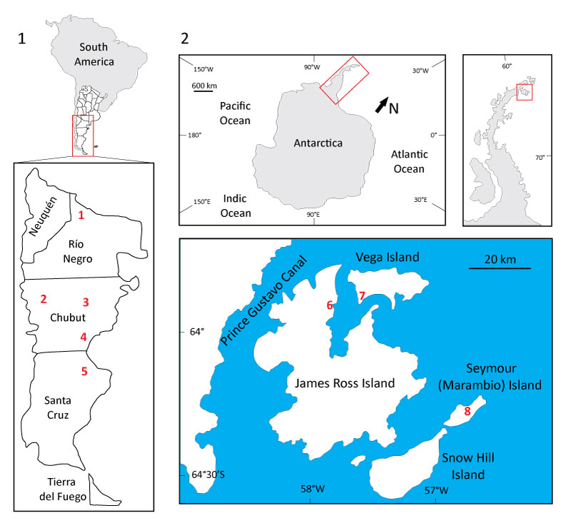
FIGURE 2. Phylogenetic relationships of dinosaurs. 1. Simplified relationships of archosaurs (adapted from Benton, 2004). 2. Phylogenetic relationships of Theropoda (adapted from Holtz et al., 2004). 3. Phylogenetic relationships of Sauropodomorpha (adapted from Yates, 2003; Pol et al., 2011;Carballido et al., 2011; D'Emic, 2012). 4. Phylogenetic relationships of Ornithischia (adapted from from Butler et al., 2008). Black circles represent node-based taxon, whereas black arrows denote stem-based taxon.
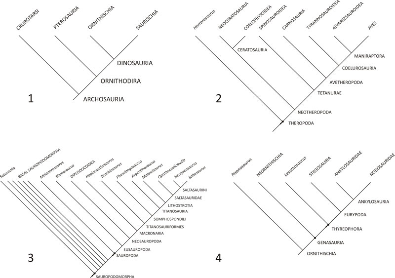
FIGURE 3. Amygdalodon patagonicus (lectotype). Posterior dorsal vertebra (MLP 46-VIII-21-1/2) in right lateral (1), anterior (2), left lateral (3), and posterior (4) views. Scale bar equals 100 mm.
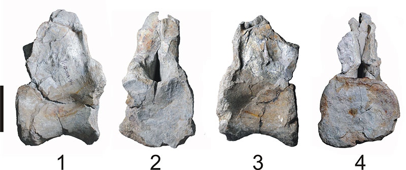
FIGURE 4. Laplatasaurus araukanicus (lectotype). Right tibia (MLP-CS 1128) in medial (1), anterior (2), lateral (3), posterior (4), and proximal (5, anterior towards top) views. Right fibula (MLP-CS 1127) in lateral (6), posterior (7), medial (8), and anterior (9) views. Scale bar equals 100 mm.
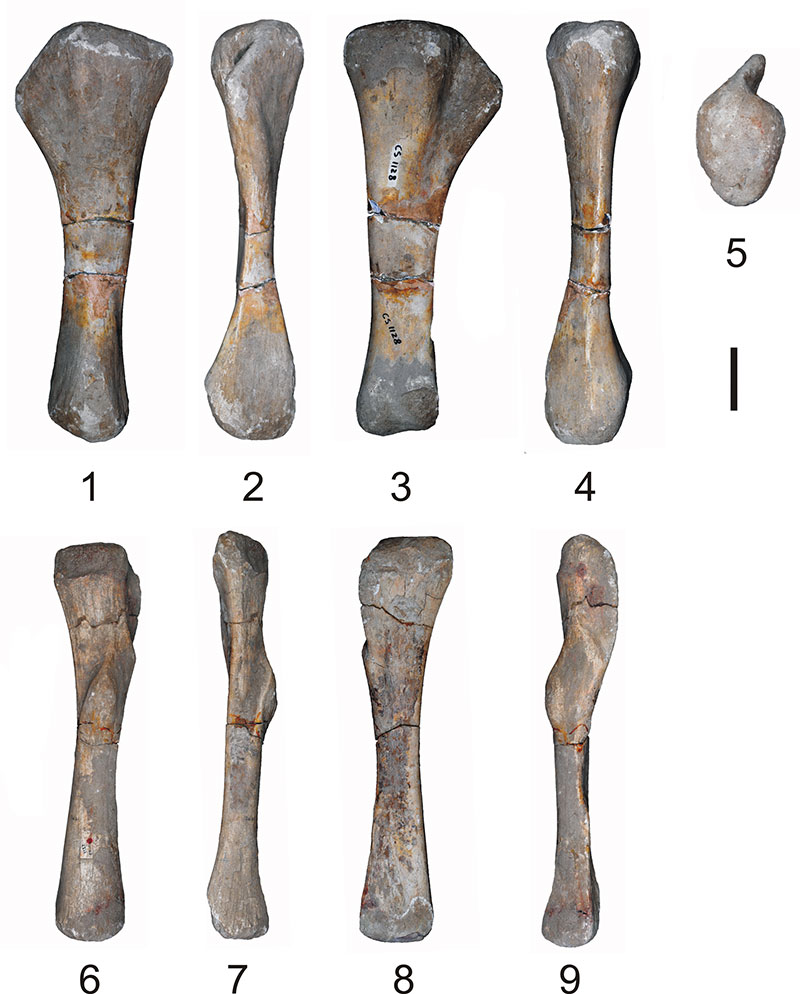
FIGURE 5. Microcoelus patagonicus nomen dubium (holotype). Anterior dorsal vertebra (MLP-Ly 23) in left lateral (1), posterior (2), and right lateral (3) views. Scale bar equals 100 mm.
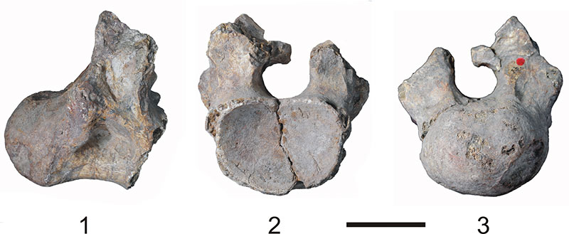
FIGURE 6. "Titanosaurus" nanus nomen dubium (holotype). Cervical vertebra (MLP-Ly 19) in right lateral (1), posterior (2), and anterior (3) views. Dorsal vertebra (MLP-Ly 18) in left lateral (4), posterior (5), and anterior (6) views. Scale bar equals 100 mm.
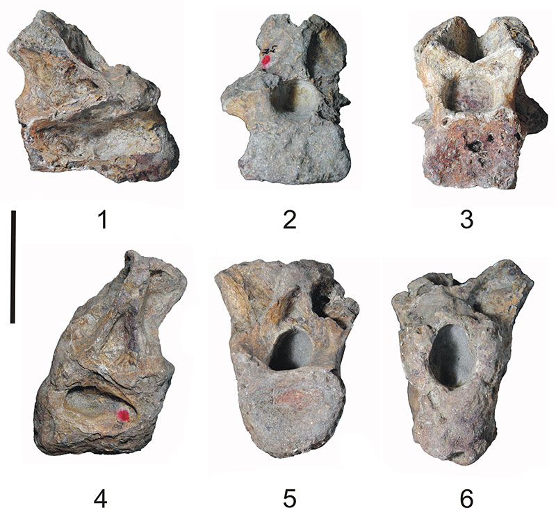
FIGURE 7. Argyrosaurus superbus, holotype (MLP 77-V-29-1). 1. Left humerus in anterior view. 2. Left ulna in anterolateral view. 3. Left radius in posterior view. 4. Left metacarpus in anterior view. Scale bar equals 100 mm.
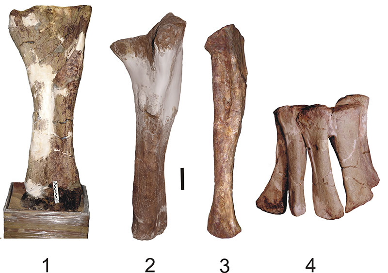
FIGURE 8. Antarctosaurus giganteus (holotype). Femora (MLP 26-316) exhibited on display at the MLP.
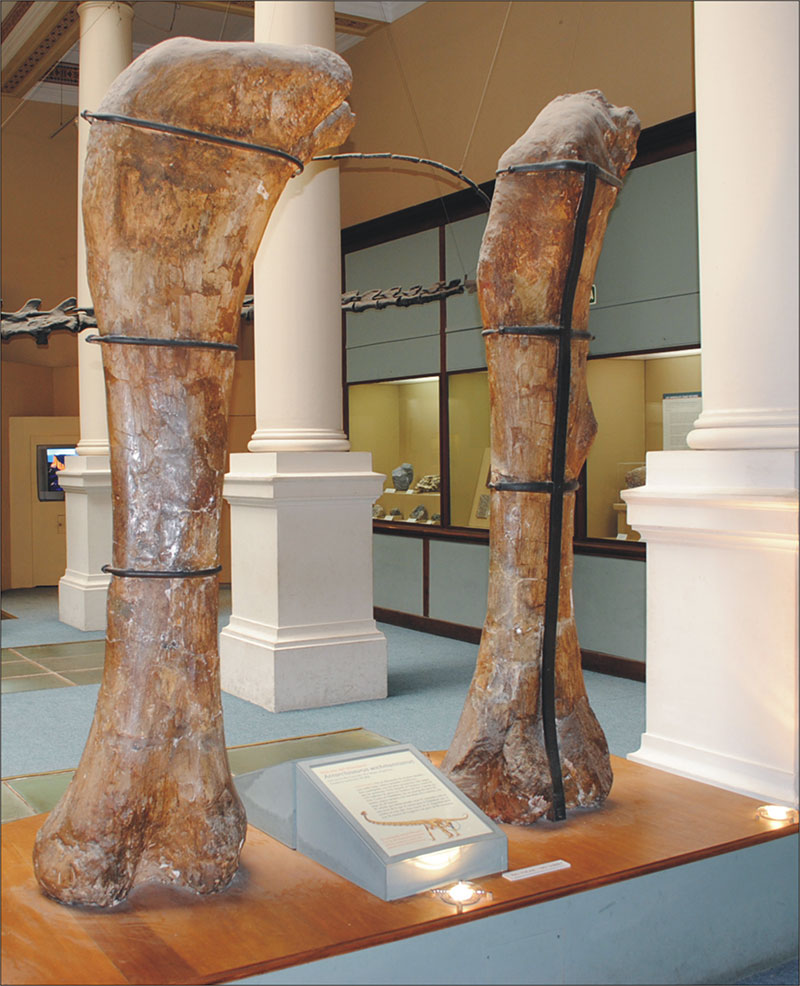
FIGURE 9. Lithostrotia gen. et sp. ideterminate. Caudal vertebrae (MLP 11-II-20-1) in anterior (1), right lateral (2) and posterior (3) views. Scale bar equals 100 mm.
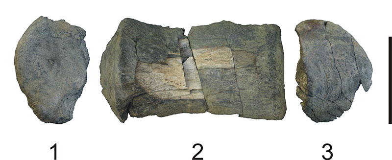
FIGURE 10. Neuquensaurus australis (holotype). 1. Sacral vertebrae 1-5 (MLP-Ly 7). 2. Sacral vertebrae 5 and 6 (MLP-Ly 7). 3. Sacral vertebra 7, biconvex (MLP-Ly 1). 4. Second caudal vertebra (MLP-Ly 2). 5. Third caudal vertebra (MLP-Ly 3). 6. Anterior caudal vertebra (MLP-Ly 4). 7. Anterior caudal vertebra (MLP-Ly 5). 8. Middle caudal vertebra (MLP-Ly 6). All elements are in right lateral view (reversed). Scale bar equals 100 mm.
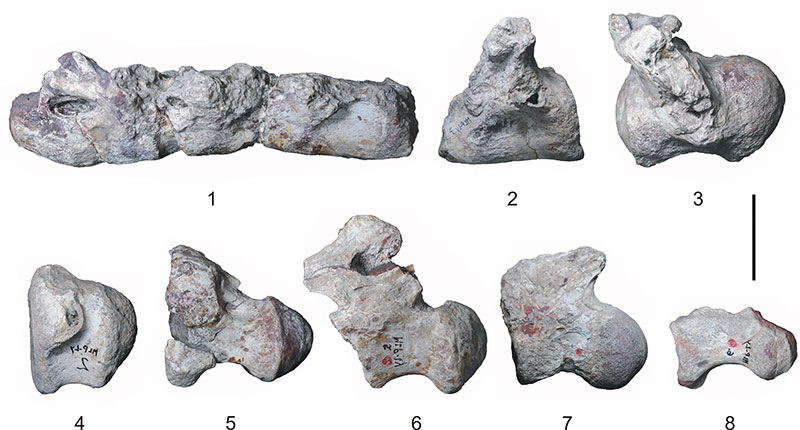
FIGURE 11. Neuquensaurus robustus (lectotype). Left femur (MLP-CS 1480) in anterior (1) and posterior (2) views. Left radius (MLP-CS 1171) in posterior (3), lateral (4), anterior (5), and medial (6) views. Left ulna (MLP-CS 1094) in lateral (7), posterolateral (8), proximal (9, anterior towards top), and anteromedial (10) views. Right ulna (MLP-CS 1095) in lateral (11), posteromedial (12), proximal (13, anterior towards top), and anteromedial (14) views. Scale bar equals 100 mm.
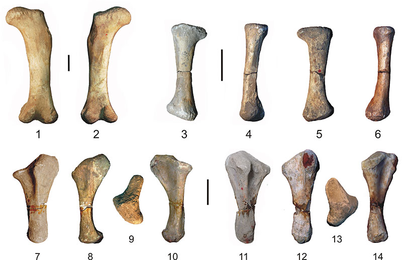
FIGURE 12. Genyodectes serus (holotype) (MLP 26-39). Premaxilae in anterior (1), posterior (2), right lateral (3) and left lateral (4) views. Right maxilla in lateral (5) and medial (6) views. Left maxilla in lateral (7) and medial (8) views. Right dentary in medial (9) and lateral (10) views. Left dentary in medial (11) and lateral (12) views. Scale bar equals 100 mm.
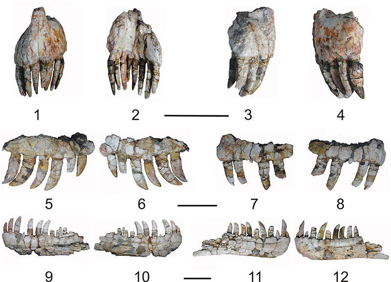
FIGURE 13. Antarctopelta oliveroi, holotype (MLP 86-X-28-1). Partial. 1. Left dentary in medial view. 2. Teeth. 3. Anterior cervical vertebra in anterior view. 4. Posterior cervical vertebra in anterior view 5. Right IV? metatarsal in ventral view. Scale bar equals 10 mm in 1 and 2, and 3 mm in 3, 4 and 5.
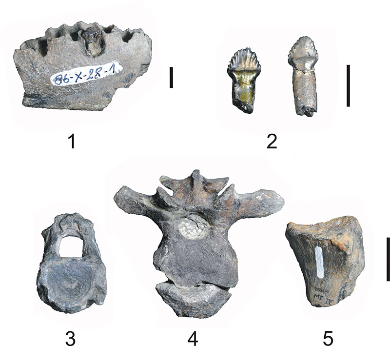
FIGURE 14. Hadrosauridae indet. (MLP 99-I-10-1). Tooth in occlusal (1) and side (2) views. Scale bar equals 10 mm. (adapted from Case et al., 2000).
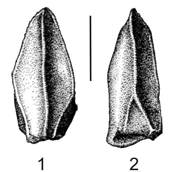
FIGURE 15. Hadrosauridae? (MLP 96-I-6-2). Distal end of metatarsal in dorsal (1), lateral or medial (2), ventral (3), lateral or medial (4), and distal (5) views. Scale bar equals 50 mm.
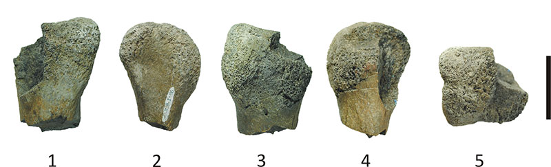
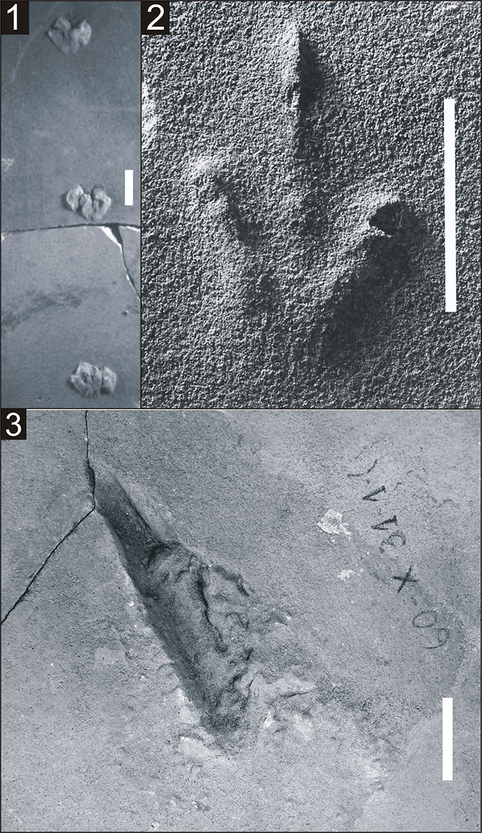
FIGURE 16. Ichnotaxa. 1. Delatorrichnus goyenechei, holotype (MLP 60-X-31-6) (adapted from De Valais, 2011). 2. Wildeichnus navesi, paratype (MLP 60-X-31-11). Sarmientichnus scagliai, holotype (MLP 60-X-31-1A). Scale bar equals 30 mm.