FIGURE 1. Spatiotemporal distributions of middle-Eocene carnivoraforms in southern California. 1, Map of localities that have yielded carnivoraforms. 2, Stratigraphic distributions of mammalian carnivores. See legend for color coding of various geologic formations. Temporal correlation of fossil assemblages, lithologic units, and magnetochrons follows Walsh (1996), Walsh et al. (1996), Mihlbachler and Deméré (2009), and Kelly et al. (2012), and generally should not be regarded as precise above one million-year resolution (cf. Walsh, 1996). See Lander (2011) for alternative age assignments for Mission Valley Formation and local faunas of Brea Canyon, Tapo Canyon, and Jeff's Discovery site. Vertical lines represent minimum age uncertainties. Significant local faunas are indicated as follows: B, Brea Canyon; J, Jeff's Discovery; L, Laguna Riviera; P, Poway; R, Pearson Ranch; T, Tapo Canyon. Original map for Figure 1.1 generated by BerkeleyMapper (http://berkeleymapper.berkeley.edu/).
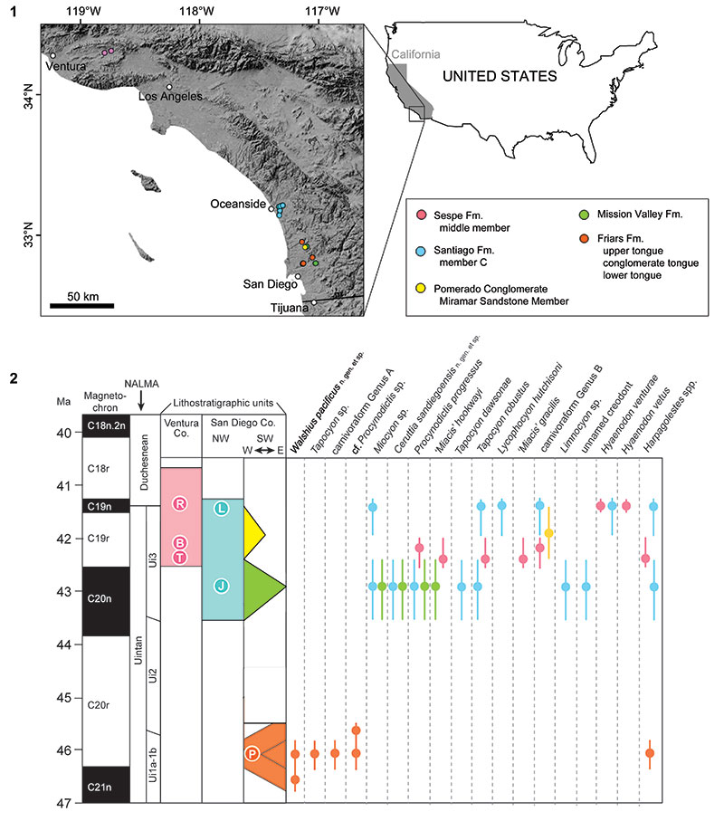
FIGURE 2. Dentition of Ceruttia sandiegoensis n. gen. et sp. Left premaxilla and maxilla of the holotype SDSNH 92504 in ventral view (1); left P4-M2 of SDSNH 92504 in occlusal view (2); left dentary fragment of SDSNH 92503 in labial view (3); left dentary fragment of SDSNH 42812 in labial (4), occlusal (5), and lingual (6) views; right dentary fragment (7) and right m2 (8) of SDSNH 42812 in occlusal view. Two white triangles in Figure 2.1 point to possible alveoli for M3. Black triangle in Figure 2.5 points to alveolus for m3. Same scale bar applies to Figure 2.1 and 2.4-2.7. Abbreviation: if, incisor foramen.
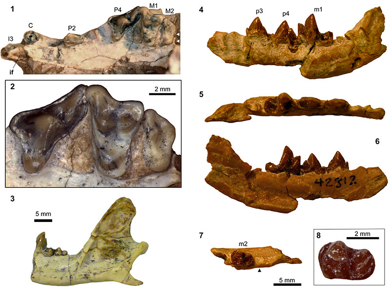
FIGURE 3. Left auditory region (SDSNH 92504) of Ceruttia sandiegoensis n. gen. et sp. Abbreviations: as, alisphenoid; er, epitympanic recess; fs, fossa for stapedius muscle; ftt, fossa for tensor tympani; fv, fenestra vestibuli; gf, glenoid fossa; Gf, Glaserian fissure; mp, mastoid process; pgf, postglenoid fossa; R, rugose area on petrosal promontorium; smf, suprameatal fossa; spa, sulcus for stapedius artery.
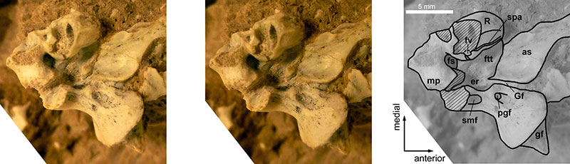
FIGURE 4. Dentition of Walshius pacificus n. gen. et sp. Right maxillary fragment of SDSNH 46197 in occlusal view (1); right maxillary fragment of SDSNH 62150 in lingual view (2, inverted);right dentary fragment of SDSNH 50599 (cf. W. pacificus) in labial (3), lingual (4), and occlusal (5) views.
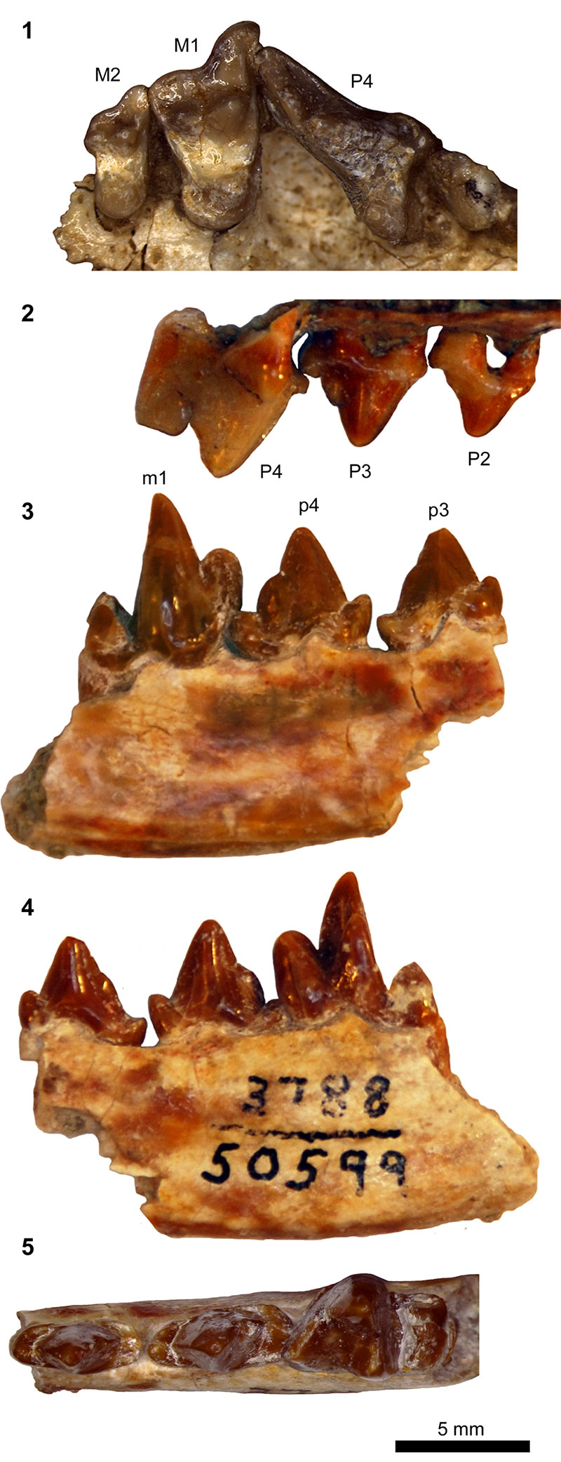
FIGURE 5. Dentition of Procynodictis and 'Miacis' gracilis. P. vulpiceps (AMNH 2514, Uinta Formation): left P3-M1 (1, 4); right dentary in labial (8) and occlusal (10) views. P. progressus (Santiago Formation): SDSNH 42813, left P4-M2 (3, 7); SDSNH 43744, right dentary in labial (9) and occlusal (11) views and left m2-m3 in occlusal view (17); SDSNH 43656, left m2 in occlusal view (16); SDSNH 42810, left dentary in labial view (18). Cf. 'M.' gracilis: LACM 40450, right P4-M1 from the Sespe Formation (2, 6; inverted). 'M.' gracilis: AMNH 104960 (cast of CM 12063) from the Uinta Formation (5). Cf. Procynodictis sp.: SDSNH 47389, left p4 in labial (12) and occlusal (14) views; SDSNH 55888, right m1 in lingual (13) and occlusal (15) views. Abbreviations: mf, mental foramina; pac, posterior accessory cuspid.
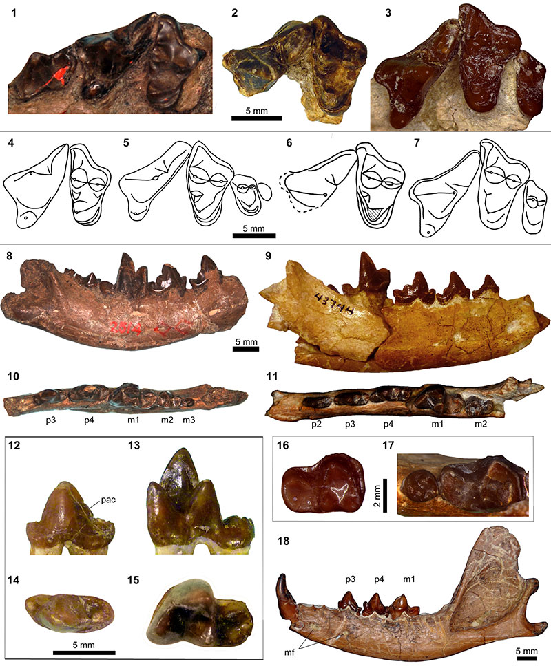
FIGURE 6. Dentition of specimens referred to 'Miacis' hookwayi. LACM 40463 (cf. 'Miacis' hookwayi), left maxillary fragment with P4-M2 (1) and right maxillary fragment with P3-M2 in occlusal (2) and lingual (3) views; LACM CIT 1656 (holotype), left dentary fragment with m1-m2 in occlusal view (4, inverted); SDSNH 84969, right m1-m2 in occlusal view (5).
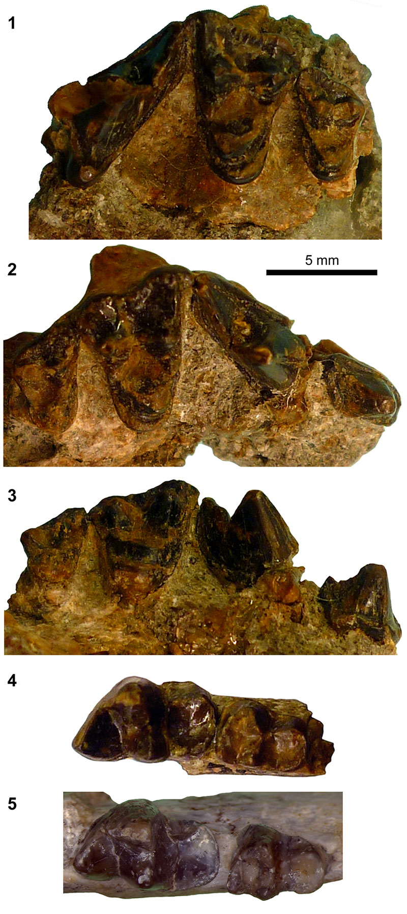
FIGURE 7. Undeterminate carnivoraform Genus A. Left dentary fragment with p4-m2 (UCMP 110072) in labial (1), lingual (2), and occlusal (3) views.
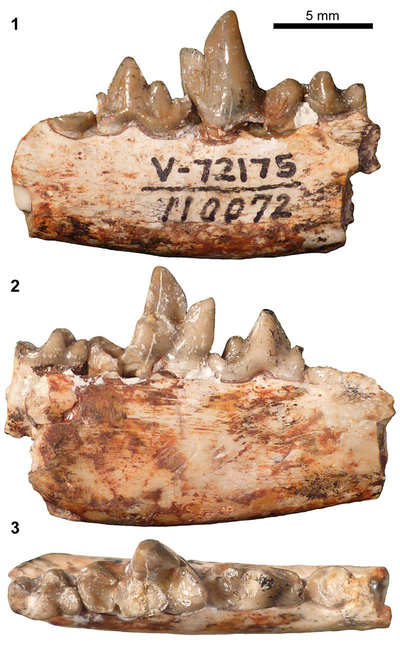
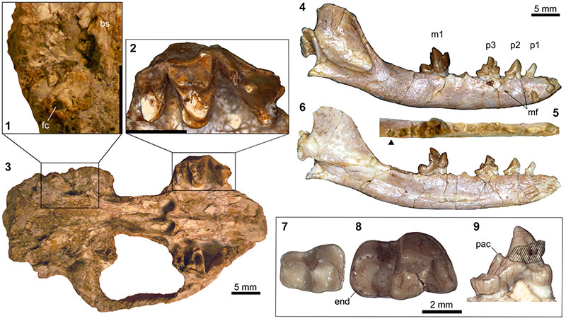
FIGURE 8. Undeterminate carnivoraform Genus B. SDSNH 56335, right middle-ear region (1), right P4-M2 (2) and cranium (3) in ventral view; UCMP 141385, left dentary in labial (4, inverted), occlusal (5), and lingual (6) views, left m1 in occlusal view (8), and left p3 in lingual view (9); UCMP 314191, left m2 in occlusal view (7). Black triangle in Figure 8.4 points to alveolus for m3. Scale bars for Figure 8.1 and 8.2 equal 5 mm. Figure 8.4-8.6 at the same scale. Abbreviations: bs, basisphenoid; end, entoconid; fc, fenestra cochlea; mf, mental foramina; pac, posterior accessory cuspid.