FIGURE 1. Location map showing the locality where Eosclerocalyptus cf. E. lineatus (Xen-30) was exhumed.
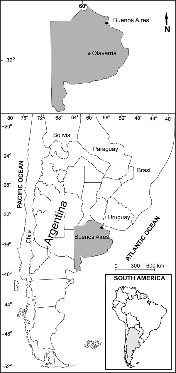
FIGURE 2. Lithostratigraphical profile at the locality where Eosclerocalyptus cf. E. lineatus (Xen-30) was exhumed.
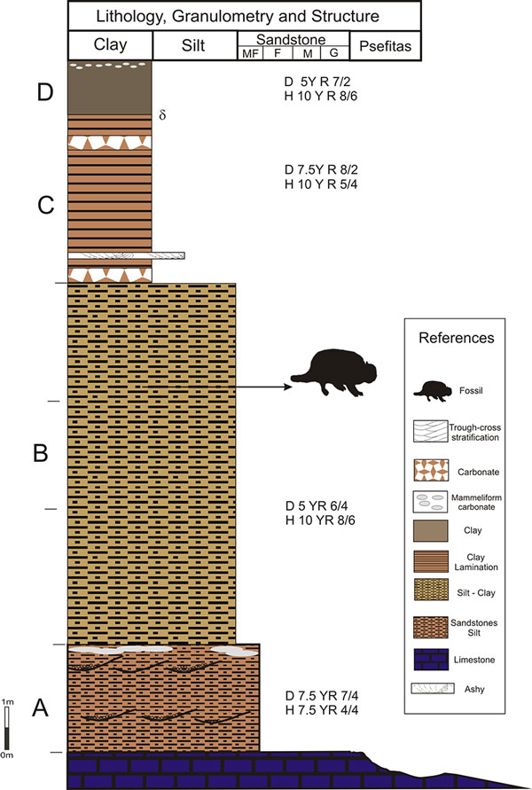
FIGURE 3. 1 to 4: Recreations of the 3D disposition in situ (from four different angles) of Eosclerocalyptus cf. E. lineatus (Xen-30). Abbreviations: a, posterior fused vertebrae; b, right hemimandible; c, anterior fused vertebrae; d, left zeugopod and autopod; e, atlas and axis; f, scapula; g, synsacrum; h, dorsal carapace.
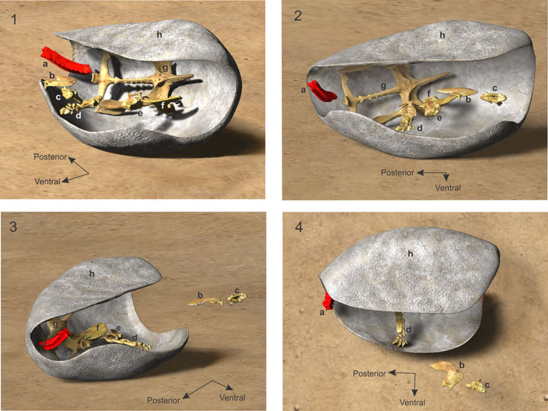
FIGURE 4. Sequence of the four episodes of the burial (numbered from 1 to 4) of Eosclerocalyptus cf. E. lineatus (Xen-30). Abbreviations: a, posterior fused vertebrae; b, right hemimandible; c, anterior fused vertebrae; d, left zeugopod and autopod; e, atlas and axis; f, scapula; g, synsacrum; h, dorsal carapace.
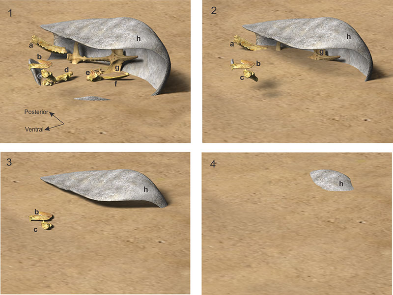
FIGURE 5. Posterior fused vertebrae of Eosclerocalyptus cf. E. lineatus (Xen-30-12) with a detail of the pits and furrows observed on the neural apophysis.
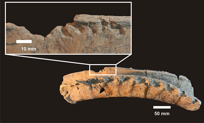
FIGURE 6. 1. Pits and furrows (i to vi) on the neural apophysis of Eosclerocalyptus cf. E. lineatus (Xen-30-12). 2. Palatal view of the rostrum of Capalmalania altifrontis (MMP-1121-M) showing incisors and canines. Abbreviations: I1 first upper incisor, I2 second upper incisor, I3 third upper incisor, C upper canine.
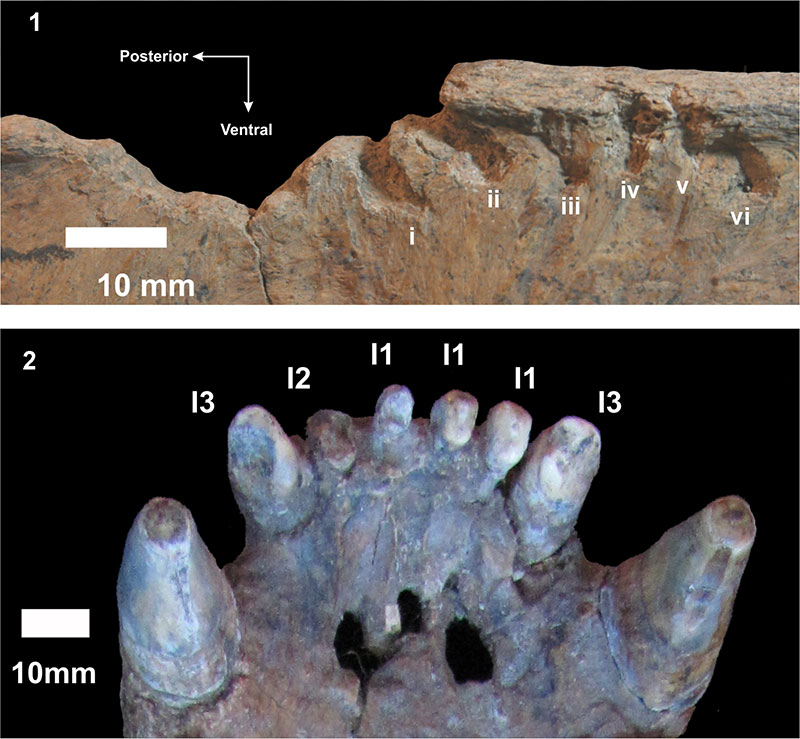
FIGURE 7. Two different views of acombined image of the rostrum of Chapalmalania altifrontis (54-V-17-1) "biting" the posterior vertebrae of Eosclerocalyptus cf. E. lineatus (Xen-30-12) showing the precise match between the dentition of Ch. altifrontis and the marks on the vertebrae. Scale bar equals 10 mm.
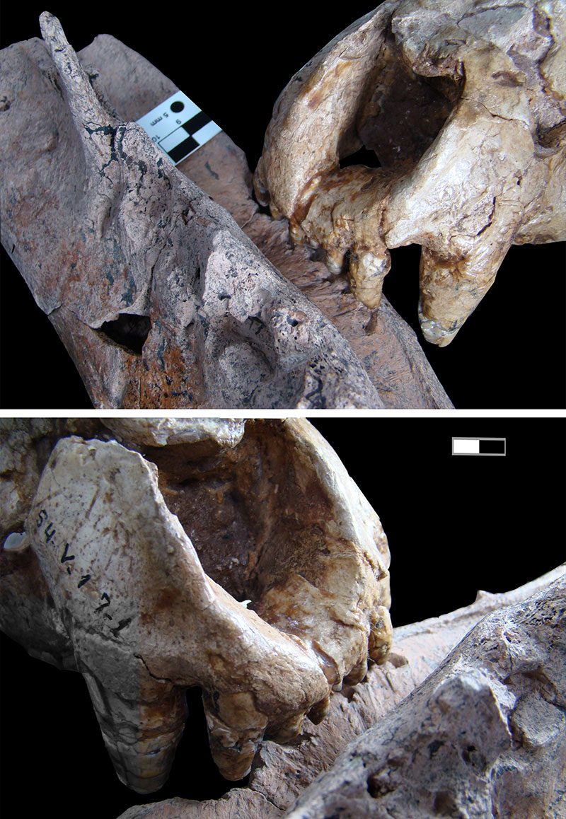
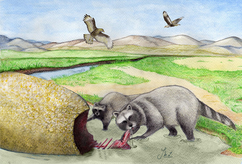
FIGURE 8. Reconstruction of the scene of consumption (By Luz Irrazábal).