FIGURE 1. 1: Upper Jurassic shell bed, Svalbard, with ammonite (Dorsoplanites sp.) and bivalves (mainly Buchia sp.). PMO 217.501. Field of view 14 cm. Virtually relighted PTM image, comparable with an unprocessed standard photograph. 2: Physically painted black, followed by ammonium chloride whitening, standard photography, some contrast stretch.
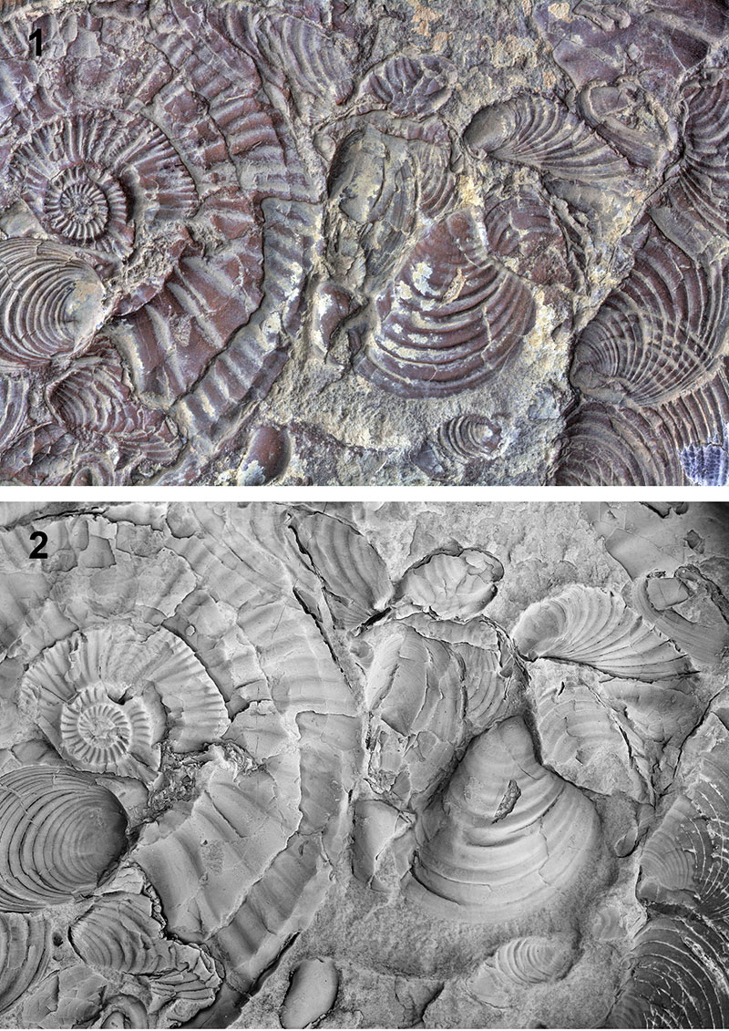
FIGURE 2. Best effort virtual whitening with Phong shading using the specular enhancement method of Malzbender et al. (2000), with low exponent value to reduce shininess.
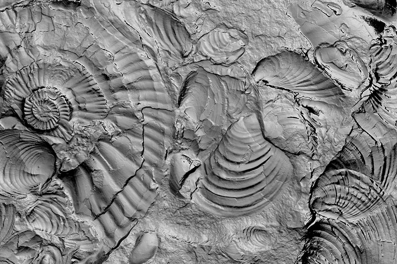
FIGURE 3. For each pixel with position (u, v) in the image plane, luminosity is modelled with a biquadratic polynomial as a function of incoming light direction. The direction is given by the projection (lu, lv) of the light vector (normalized to unit length) onto the image plane.
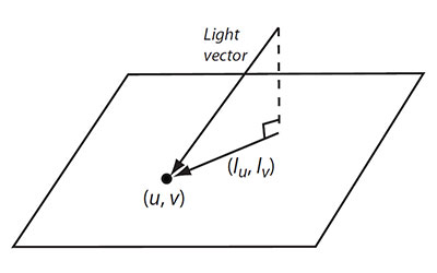
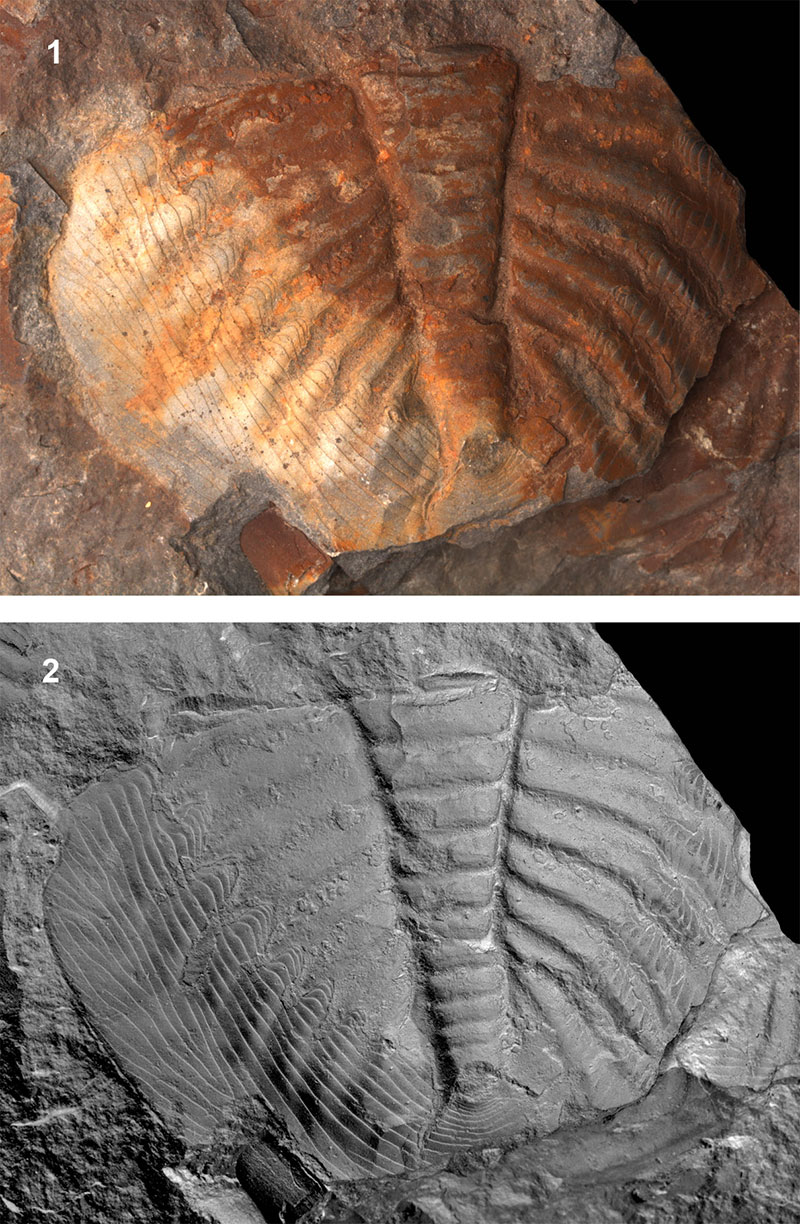
FIGURE 4. Virtual whitening of a trilobite (Bathyuriscus sp., Middle Cambrian, British Columbia), length 3 cm. 1: Virtually relighted grayscale PTM image, no further processing. 2: Virtually whitened. 3: Virtually whitened, with black paint effect. Note the darkening of concave features, e.g., on the pleural furrows.
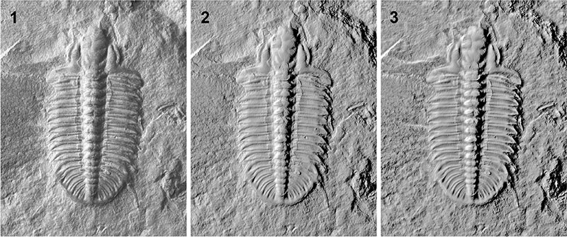
FIGURE 5. 1: Virtual whitening of an Upper Jurassic shell bed, cf. Figures 1-2. 2: Virtually whitened, with black paint effect.
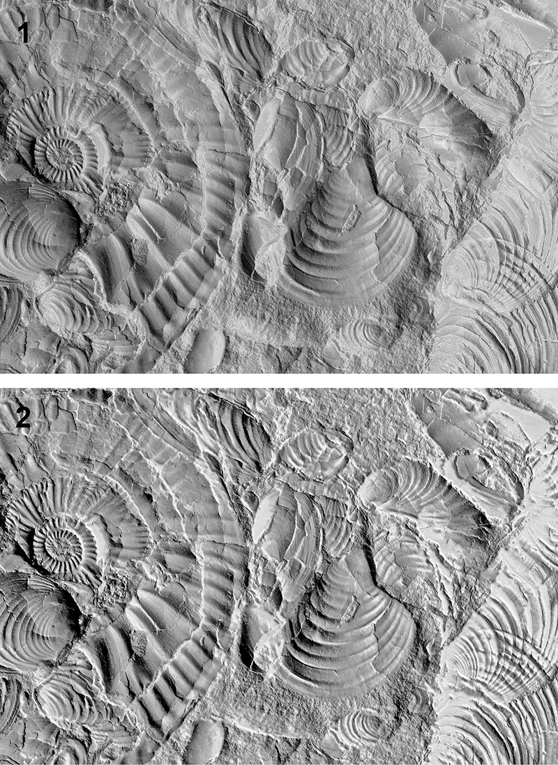
FIGURE 6. Virtual whitening of an Upper Jurassic mega-onychite (4 cm long), Svalbard. PMO 223.379. 1: Original image. 2: Virtually whitened, without black paint effect.
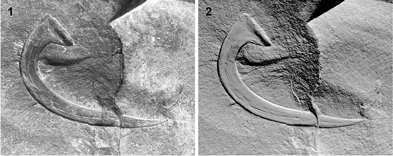
FIGURE 7. Virtual whitening of a Middle Ordovician trilobite pygidium (Ogygiocaris, Oslo Region, PMO 60422). 1: Original image. 2: Virtually whitened, without black paint effect. The sharp colour transition in the original has been completely removed, and terrace lines are greatly enhanced.