FIGURE 1. 1, Geographical setting of the collection site, a roadcut on the eastern side of the Salamón-Lois road (León, NW Spain). 2, Geological sketch map of the Cantabrian Zone showing the location of the Lois-Ciguera sector (modified from Pérez-Estaún et al., 1988).
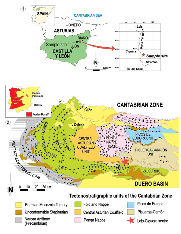
FIGURE 2. Schematic section of the Bachende Formation showing the stratigraphic position of the type locality.
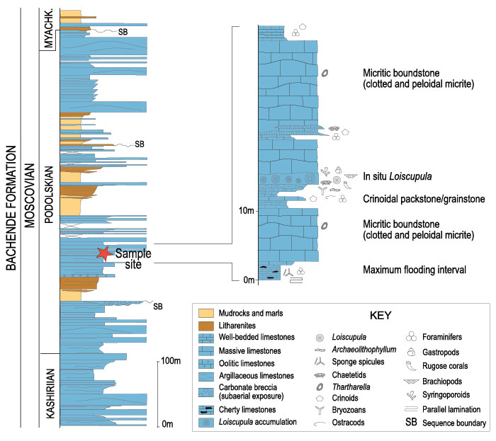
FIGURE 3. General morphologic features of Loiscupula bachendensi gen. nov. sp. nov. from the Bachende Formation , Cantabrian Zone, NW Spain. 1, type locality of Loiscupula bachendensi in the Bachende Formation, arrows point to different specimens. 2-6 Holotype USAL LB1, 2, side view showing the flat cup-shaped skeleton, with the central part of the specimen lower than the outer edges. 3, top view, showing the wackestone matrix filling the cup. 4, underside of the sponge, showing the plate-shape and the concentric rings around a central point; note the projected structures developed within each ring and usually preserved as eroded (truncated) protuberances (arrow). 5, enlargement of the rectangular area in Figure 3.4, showing in detail one of the projections on the underside of the specimen. 6, close-up of the undulating underside of the skeleton, showing the eroded protuberances and encrusting bryozoans; B= bryozoans, Bq= brachiopod. 7-8, Paratype USAL LB5, 7, cylindrical feature (rectangle) growing up from the upper surface of the skeleton of Loiscupula (white arrows); B= brachiopod. 8, close-up of the rectangular area in Figure 3.7; arrows point to the poorly preserved microstructure of Loiscupula. Note the abundant foralgal encrustations (E); T= Tuberitina; Sp= sparry calcite.
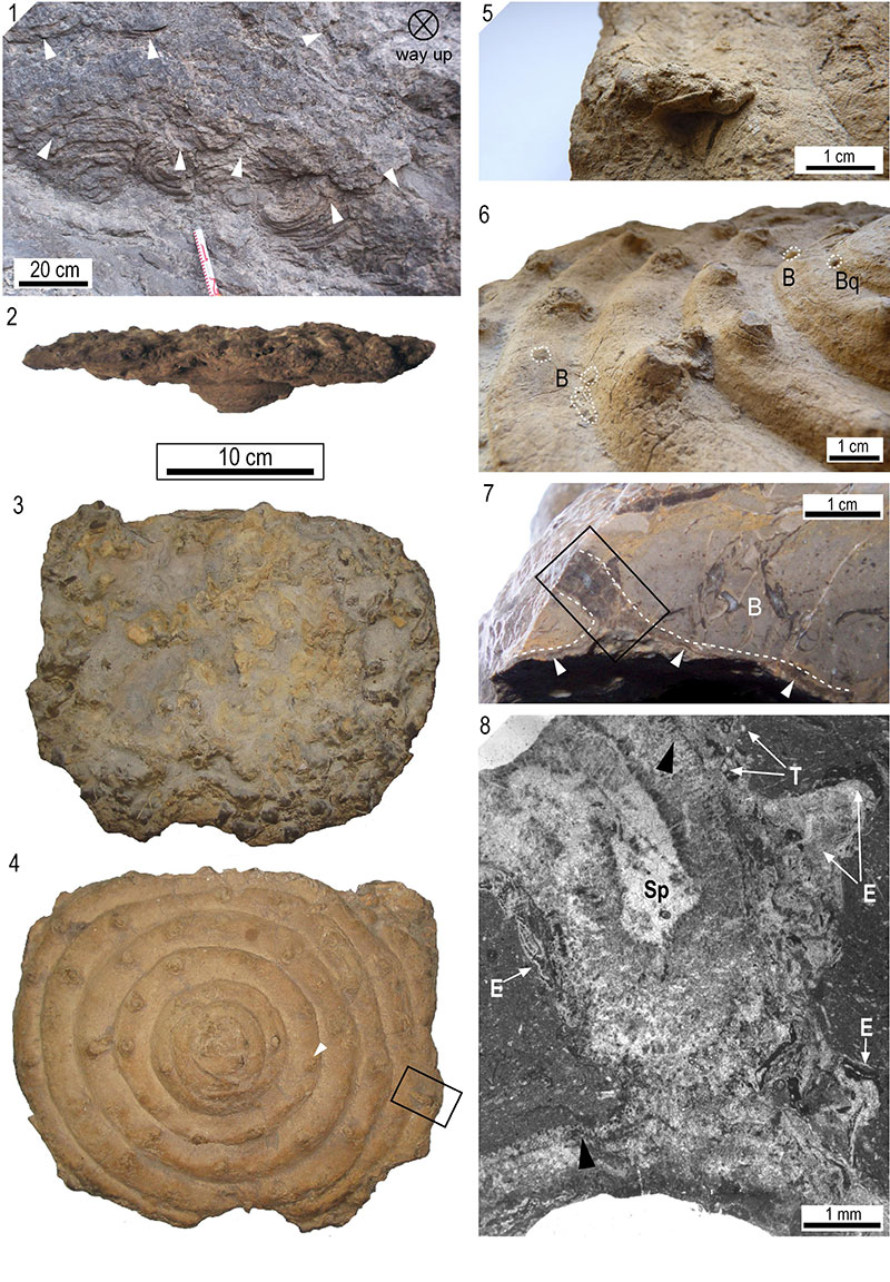
FIGURE 4. Internal structure of Loiscupula bachendensi gen. nov. sp. nov. 1, paratype USAL LB22, close-up showing the honey-comb pattern of the surface of Loiscupula bachendensi. 2, paratype USAL LB5, internal structure of the plate-like basal skeleton (enclosed by white dashed lines); white arrows point to vertical growth interruptions of the tubules; O= Ozawainella; P= pyrite. 3-4, paratype USAL LB29, 3, photomicrograph showing the internal structure of a cylindrical feature developed on the upper surface of the skeleton. 4, close-up of tubules at the upper edge of the skeleton. 5-6, Holotype USAL LB1, 5, transversal section of a cylindrical form; note the internal cavity, filled-up with micritic matrix, and the internal structure of Loiscupula composed of tubules. 6, close-up of the rectangular area in Figure 4.5 (rotated 45º counterclockwise); note the increasing of new tubules by intertubule increase (white arrow) and their horizontal partitions at different levels (T= tabulae). 7-8, paratype USAL LB5, 7, plane polarized light photomicrograph showing the overall recrystallization of the basal skeleton. 8, cathodoluminescence image of same field of view as Figure 4.7, showing the internal structure of the skeleton with development of poorly defined tabulae (arrowed).
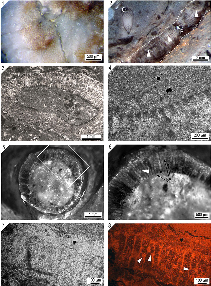
FIGURE 5. 1-8, Paratype USAL LB 5, spicule pseudomorphs of Loiscupula bachendensi gen. nov. sp. nov. All the images, except Figure 5.3, are cathodoluminescence photomicrographs. 1, Oxea (O) spicule pseudomorph floating in the surrounding matrix; white arrow is pointing to an (algal?) encrustation on the upper surface of the skeleton; T= tabulae; L=laminae, indicated by white dashed lines; note that tubules may be continuous or discontinuous across the growth interruptions. 2, intraskeletal non-luminescent spicule (mostly oxeas) pseudomorphs in a fragment of Loiscupula; white dashed line indicates the outer edge of the basal skeleton of Loiscupula; M= micritic matrix. 3, 4, plane polarized light and cathodoluminescence photomicrographs showing the internal structure of the plate-like basal skeleton; C= void filling cement; L= Loiscupula; M= micritic matrix. 5, close-up of the rectangular area in Figure 5.4, showing in detail a polyactine (P) and pseudomorphs of style spicules (S) embedded in the basal skeleton. 6, Oxea (O) and reduced polyaxon (P) spicule pseudomorphs and a probable pseudomorph of tylostyle spicule (T); dashed line indicates a fragment of Loiscupula. Note the dull appearance of the surrounding matrix and the bright to yellow-dull CL of the skeletal calcite. 7, Spicule pseudomophs concentrated towards the upper edge of the basal skeleton; dashed line indicates a transversal section of Loiscupula; T= tabula. 8, Close-up view of the rectangular area in Figure 5.5; arrow points to spicule pseudomorphs projecting into a tubule.
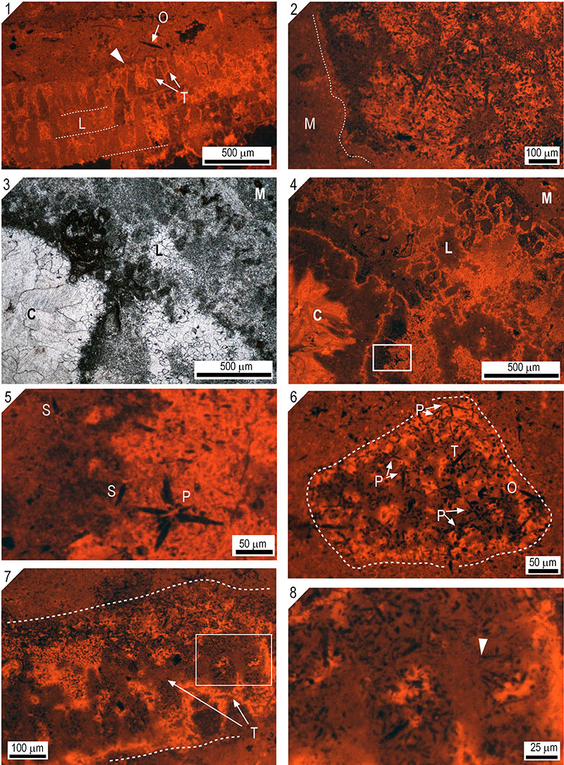
FIGURE 6. 1-6, Paratype USAL LB 5, spicule pseudomorphs and microstructure of Loiscupula bachendensi gen. nov. sp. nov. All the images are cathodoluminescence photomicrographs, except Figure 6.6, which is a scanning electron micrograph. 1, strongly recrystallized fragment of Loiscupula (L) floating in a microspartic matrix (M). 2, cathodoluminescence image of same field of view as Figure 6.1, showing abundant spicule pseudomorphs embedded in the basal skeleton; note that they are absent in the surrounding matrix; L= Loiscupula; M= matrix. 3, close-up of the rectangular area in Figure 6.2, showing in detail several pseudomorphs of oxea pseudomorphs; arrows point to spicule cross sections. 4, 5, plane polarized light and cathodoluminescence photomicrographs showing a Tuberitina specimen (T) with well developed cathodoluminescence zonation; (P) polyaxon spicule pseudomorph floating in the surrounding microsparitic matrix with some rays shortened an others missing (arrows); O= pseudomorphs of oxea spicules with irregular edges. 6, Relic fascicular penicillate microstructure of Loiscupula, composed of fibers, inferred to be aragonite needles (white arrows) within coarse calcite crystals (black arrows).
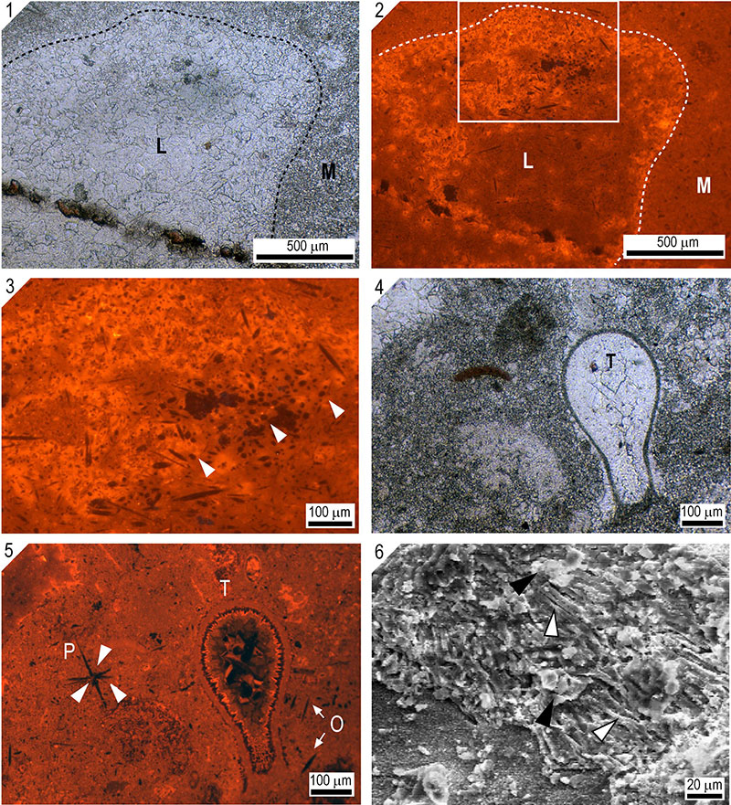
FIGURE 7. 1, Paratype USAL LB 5, Loiscupula (L) fragments floating in a muddy matrix; Ls= lasiodiscids; C=cyanobacteria filaments (Girvanella?); S= Shamovella. 2-4, USAL LB34, 2, polished slab showing the matrix that filled the cup-like skeleton of Loisphyllum (arrowed), with a complex intergrowth of Archaeolithophyllum lamellosum; note the undulating morphology of the Loiscupula skeleton (white dashed lines), representing the circular and concentring rings (grooves and ridges) in transverse section. 3, Archaeolithophyllum thallus showing relics of coaxial perithallum and hypotallum organization. 4, close-up view of the rectangular area in Figure 6.2; note the neomorphic calcite forming the basal skeleton and the poor preservation of the tubules (arrow); A= blade fragments of Archaeolithophyllum. 5, paratype USAL LB38-1, polished slab of a Loiscupula bachendensi specimen (black arrow and dashed line) with a fistuliporid bryozoan encrusting (lower left of the image) the underside of the specimen; note the development of the complex intergrowth of Archaeolithophyllum lamellosum (white arrows) in the wackestone matrix and the encrustations of small foraminifers (F), usually developed on the upper surface of the phylloid blades and Loiscupula. T= small specimen of Tetrataxis. 6, paratype USAL LB24, transversal section of a small specimen of Loiscupula bachendensi, showing the internal wackestone matrix filling the cup-shaped skeleton (dashed lines).
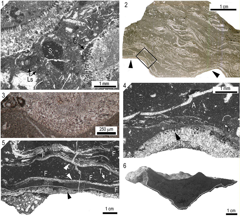
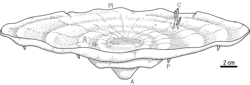
FIGURE 8. Idealized reconstruction of Loiscupula bachendensi gen. nov. sp. nov. Note the low conical growth form; A= presumed attachment point; P= projections on lower surface; G= grooves; R= ridges; Pl= plate-like skeleton; C)= cylindrical/branching features (extended mamelons/chimneys) on upper surface.