FIGURE 1. Location of the Middle Pleistocene Qesem Cave.
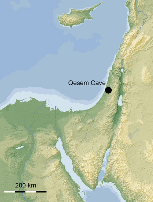
FIGURE 2. Squares of the excavation area of the Middle Pleistocene Qesem Cave with concentration (green areas) of microvertebrate fossils.

FIGURE 3. Remains of (3.1-6) Rhinolophus mehelyi Matschie, 1901, (3.7-12) Rhinolophus euryale Blasius, 1853, and (3.13-24) Rhinolophus ferrumequinum (Schreber, 1774) from the Middle Pleistocene Qesem Cave: (3.1-3) TAU-QC/CHIR-12b, left mandibular fragment, occlusal, buccal, lingual view; (3.4-6) TAU-QC/CHIR-25, left mandibular fragment, occlusal, buccal, lingual view; (3.7-9) TAU-QC/CHIR-37, right upper canine, mesial, lingual, buccal view; (3.10-12) TAU-QC/CHIR-03, right mandibular fragment, occlusal, buccal, lingual view; (3.13-15) TAU-QC/CHIR-10, right upper canine, mesial, lingual, buccal view; (3.16-18), distal epiphysis of right humerus, TAU-QC/CHIR-05, internal, posterior, external view; (3.19-21) TAU-QC/CHIR-12a and (3.22-24) TAU-QC/CHIR-46, left mandibular fragments, occlusal, lingual, buccal view; .
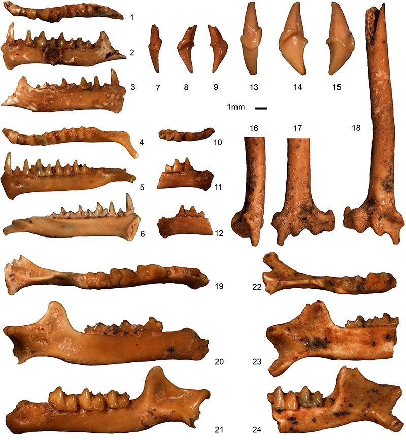
FIGURE 4. Remains of (4.1-9) Miniopterus cf. schreibersii (Kuhl, 1817), and (4.10-15) Myotis blythii (Tomes, 1857) from the Middle Pleistocene Qesem Cave: (4.1-3) TAU-QC/CHIR-04, right mandibular fragment, occlusal, buccal, lingual view; (4.4-6) TAU-QC/CHIR-07, left mandibular fragment, occlusal, buccal, lingual view; (4.7-9) TAU-QC/CHIR-48, left mandibular fragment, occlusal, lingual, buccal view; (4.10-12) TAU-QC/CHIR-40, left mandibular fragment, occlusal, buccal, lingual view; (4.13-15) TAU-QC/CHIR-02, left mandibular fragment, occlusal, buccal, lingual view.
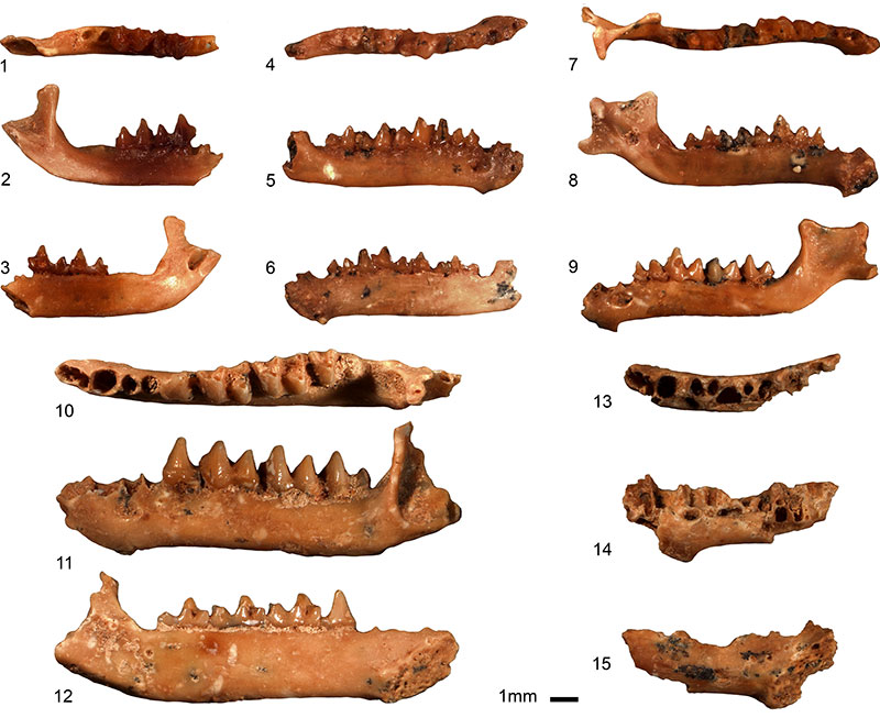
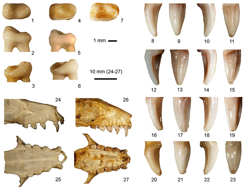
FIGURE 5. Remains of cf. Rousettus sp. from the Middle Pleistocene Qesem Cave: (5.1-3) TAU-QC/CHIR-26: right ?M1 fragment, occlusal, buccal, lingual view; (5.4-7) TAU-QC/CHIR-51: left C fragment, buccal, mesial, palatal, distal view.
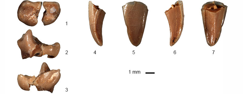
FIGURE 6. Extant Rousettus aegyptiacus (Qualamun, Dakhla, Egypt, Coll. Univ. Praha): (6.1 –7) M1 and (6.8–23) C. The views correspond to those of the fossil specimens figured in Figure 5. Upper dentitions of extant (6.24–25) Eidolon helvum (Malawi, Coll. Univ. Ceske Budejovice) and (6.26–27) Rousettus aegyptiacus (Rash130, Dakhla, Egypt, Coll. Univ. Praha), lateral and palatal views.