FIGURE 1. 1. A map showing Africa, star indicates approximate location of Lake Victoria, Rusinga Island and Mfangano Island. 2. Generalized map of Rusinga Island including basic stratigraphic distributions and general site locations. Star indicates the approximate location of this studies location. GPS coordinates for the study site: S 00° 24.350' E 034° 8.834'. 3. Generalized Miocene stratigraphy on Rusinga Island. Star indicates stratigraphic position of fossil leaf locality. Mbr. = member, Fm. = formation.
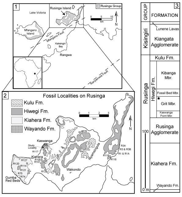
FIGURE 2. Stratigraphic section of the Grit Member exposed at fossil leaf locality. Ripple marks were found in GM-05, salt hoppers in GM-03, and fossil leaves in GM-02. GM = Grit Member.
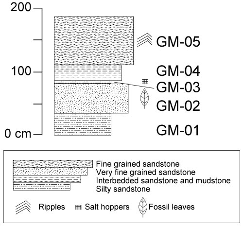
FIGURE 3. 1. Ripple marks a top GM-05. 2. Salt hoppers from GM-03. Scale bar = 1 cm.
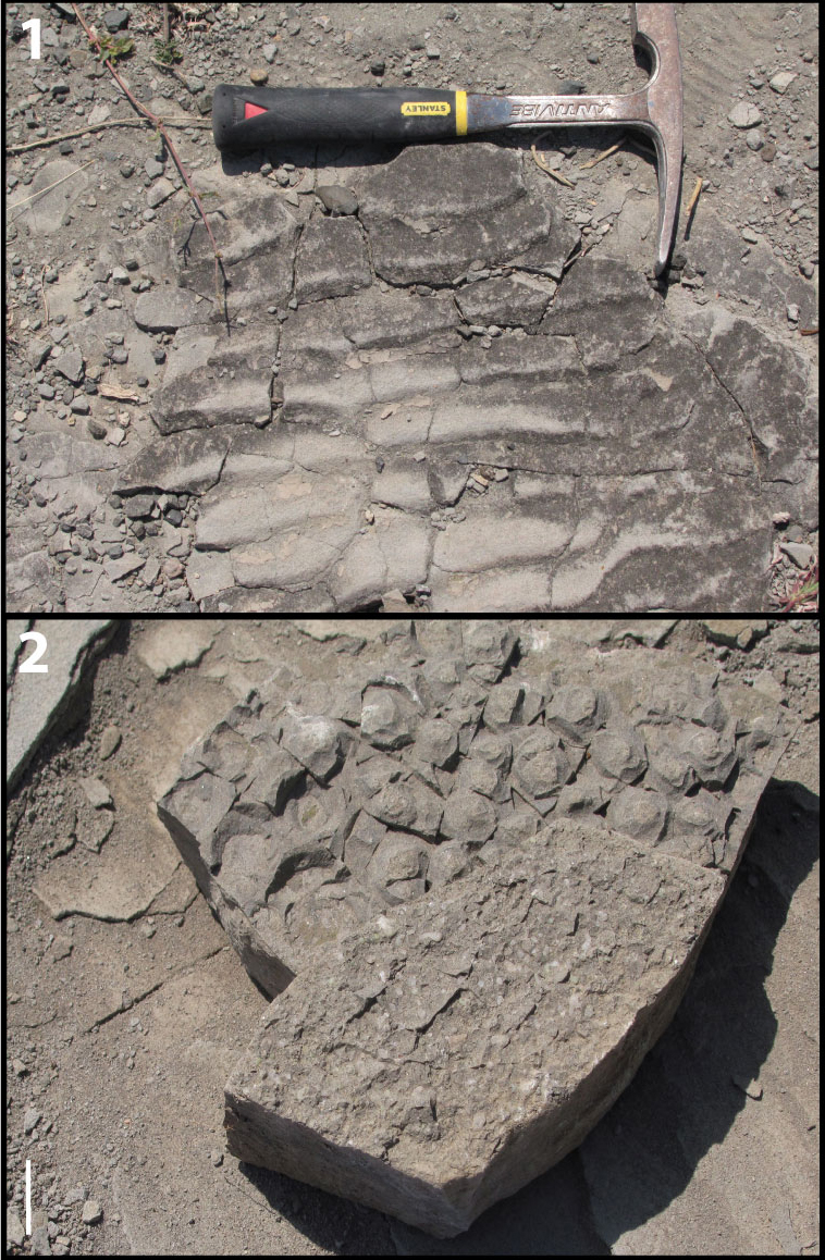
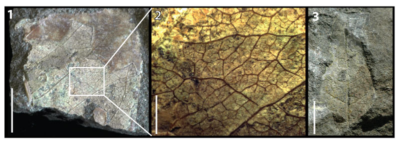
FIGURE 4. 1. Morphotype exemplar for KP-01, RU-2010-849. 2. RU-2010-864. 3. RU-2010-853. All specimens in 4.1-4.3 belong to KP-01 and display brochidodromous secondary venation, elliptic laminar shape, and cuneate base shape. 4. KP-02 morphotype exemplar, RU-2010-838, leaf showing oblong laminar shape, cordate base, pinnate primary venation, and brochidodromous secondary venation. All scales in 4.1-4.4 = 1 cm. 5. Enlarged portion of 4.4 showing cordate base with a naked basal vein, at least four basal veins, and brochidodromous secondary venation. Scale = 2 mm.
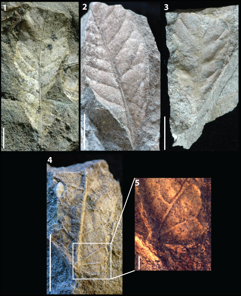
FIGURE 5. 1. KP-03 morphotype exemplar, RU-2010-267, showing eucamptodromous secondaries, highly ascending secondary angle, and abundant intersecondaries. Scale bar = 1 cm. 2. Enlarged portion of 5.1 showing straight opposite percurrent intercostal tertiaries and regular reticulate quaternary vein fabric. Scale = 2 mm. 3. RU-2010-839, leaf shows the diagnostic highly ascending secondaries unique to KP-03 along with the ovate laminar shape and straight apex shape. Scale bar = 1 cm. 4. Enlarged portion of 5.3 showing major secondaries becoming brochidodromous distally. Scale = 2 mm. 5. KP-04 morphotype exemplar, RU-2010-840, showing eucamptodromous secondary venation, low angle of divergence of secondaries that turn up abruptly near margin, and cordate base shape. Scale = 1 cm. 6. RU-2010-842, further showing the diagnostic secondary vein course and cordate base shape characteristic of KP-04. Scale = 1 cm.
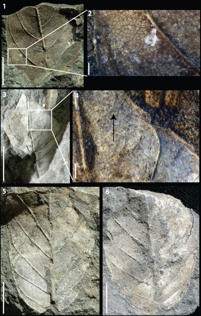
FIGURE 6. 1. KP-05 morphotype exemplar, RU-2010-844, view of whole leaf showing ovate to elliptic shape, pinnate primary venation with a convex to rounded base. Note that the leaf is slightly folded on underlying rock, causing deformation of the overall shape in photo. 2. RU-2010-844, right side of leaf showing brochidodromous secondary venation. 3. Line drawing showing brochidodromous secondary venation, simple agrophic veins, intersecondaries, and opposite sinuous percurrent tertiaries with obtuse angles. All scale bars = 1 cm.
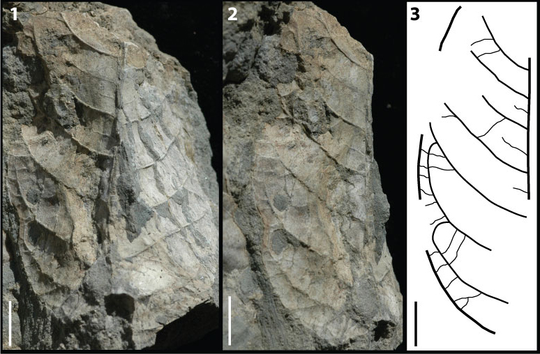
FIGURE 7. 1. KP-06 morphotype exemplar, RU-2010-845, leaf showing brochidodromous secondary venation, oblong laminar shape, and convex base shape. 2. Line drawing showing brochidodromous secondary venation, intersecondaries, and opposite sinuous percurrent tertiaries with angles varying from perpendicular to obtuse. 3. RU-2010-862, basal end of leaf showing convex base shape and brochidodromous secondary venation. 4. RU-2010-862, apical end of leaf showing acute apex angle. All scale bars = 1 cm.
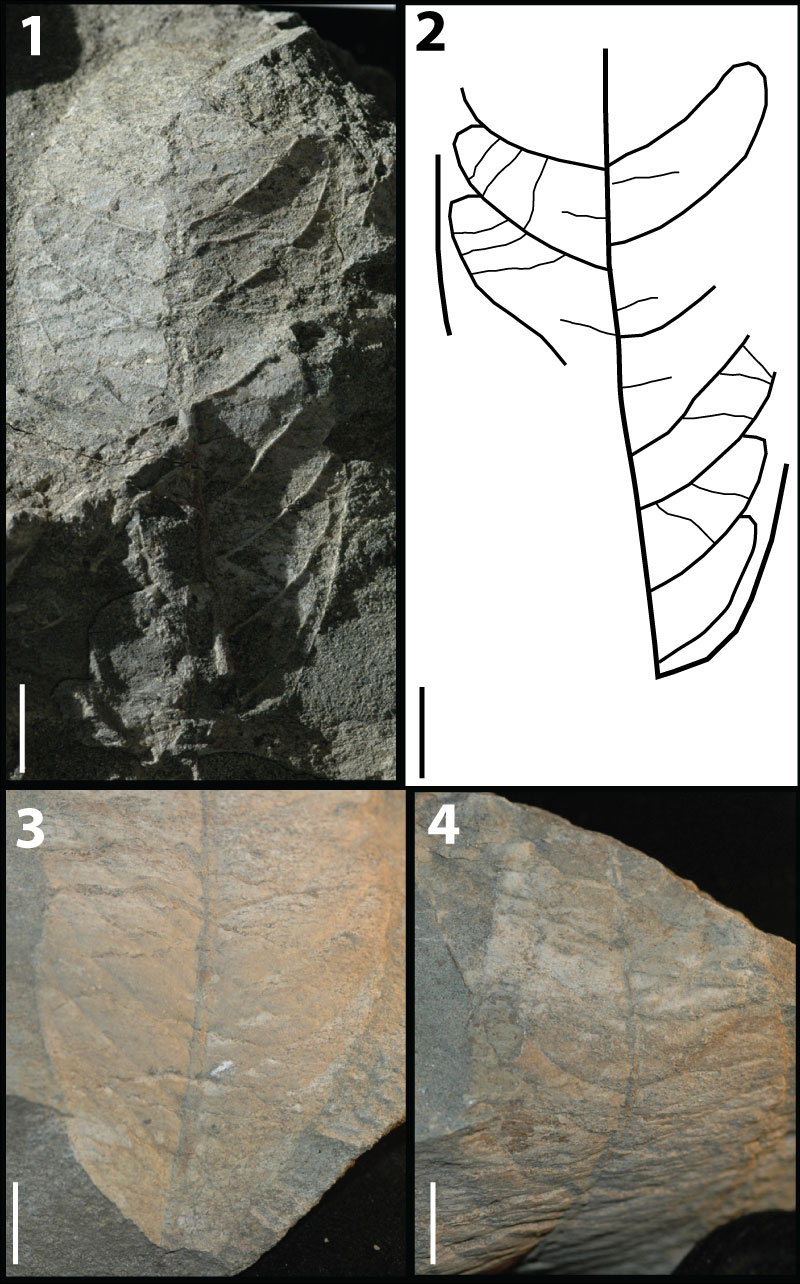
FIGURE 8. 1. KP-07 morphotype exemplar, RU-2010-848, showing ovate laminar shape, concave base shape, pinnate primary venation, and brochidodromous secondary venation. 2. Line drawing showing brochidodromous secondary venation, intersecondaries, and mixed percurrent intercostal tertiaries. 3. KP-08 morphotype exemplar, RU-2010-860, showing elliptic to oblong laminar shape, straight apex shape, and weak brochidodromous secondaries with close, uniform, spacing. All scale bars = 1 cm.
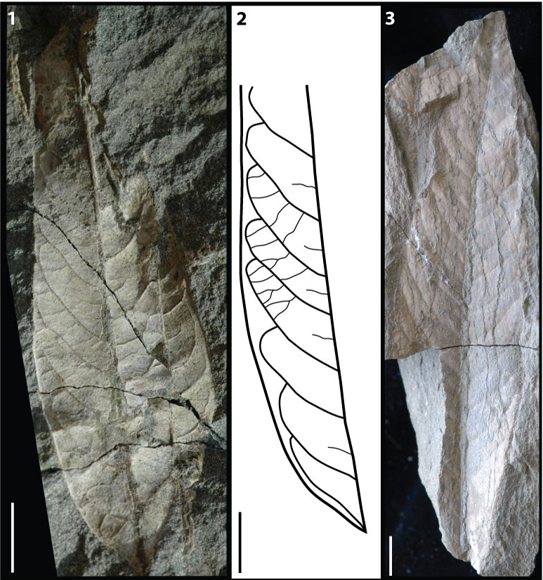
FIGURE 9. 1. KP-09 morphotype exemplar, RU-2010-850, showing ovate laminar shape, straight apex and pinnate primary venation. 2. KP-10 morphotype exemplar, RU-2010-859, showing brochidodromous secondaries with uniform angles and spacing. 3. Counterpart to RU-2010-859 showing acute apex angle and acuminate apex shape. 4. KP-11 morphotype exemplar, RU-2010-861, showing convex base shape and pinnate primary venation. 5. Enlarged portion of 9.4 showing irregular reticulate intercostal tertiaries. Scale = 2 mm. 6. KP-12 morphotype exemplar, RU-2010-863, showing regularly spaced secondaries diverging at a low angle, and a stout midvein. 7. Enlarged portion of 9.6 showing mixed percurrent intercostal tertiaries. 8. Line drawing highlighting the uniform obtuse angles of the intercostal tertiaries. All scales in 9.1-9.4 and 9.6-9.8 = 1 cm.
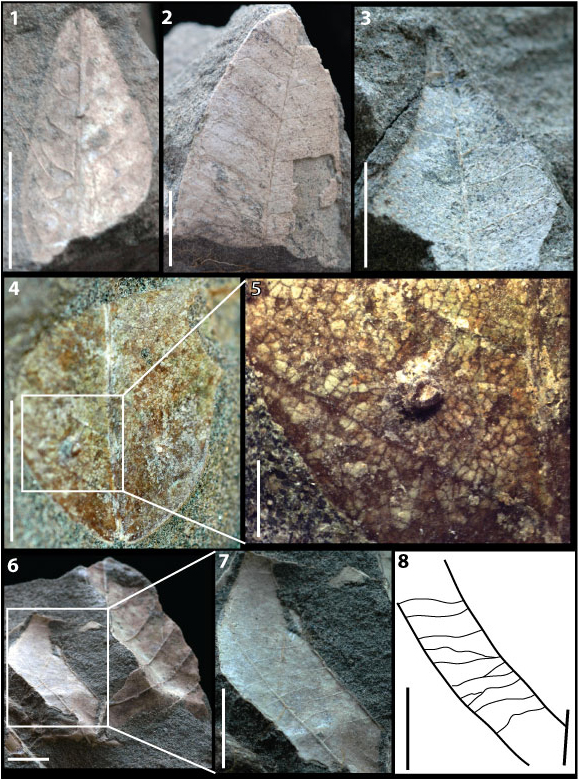
FIGURE 10. 1. KP-13 morphotype exemplar, RU-2010-866, aff. Typha sp. showing major linear veins parallel and evenly spaced. Scale = 1 cm. 2. aff. Typha sp. close up showing minor linear veins running parallel and evenly spaced between major linear veins. Scale = 2 mm. 3. KP-14 morphotype exemplar, RU-2010-867, aff. Phragmites sp. showing parallel major linear veins with midrib. Scale =1 cm. 4. Additional specimen of KP-14, aff. Phragmites sp., to further demonstrate presence of a midrib, distinguishing KP-14 from KP-13.
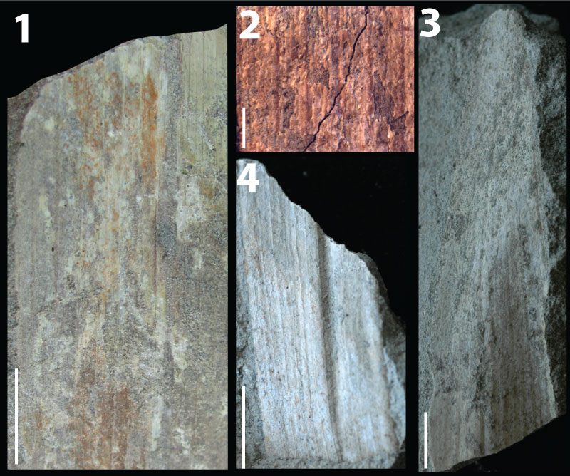
FIGURE 11. 1. KP-15 distinct dicotyledonous fragment, RU-2010-837, showing brochidodromous secondary venation and well preserved higher order venation. Scale bar = 1 cm. 2. Enlarged portion of 11.1 showing intercostal tertiaries irregular reticulate, epimedial tertiaries reticulate, exterior tertiaries variable, and quaternary vein fabric irregular reticulate. 3. KP-16, distinct dicotyledonous fragment, RU-2010-847, showing cladodromous secondary vein course and potential ovate laminar shape. This fragment is potentially the apex of a larger leaf.