APPENDIX
FIGURE A1. Photomicrographs of Protocinctus under cathodoluminescence (a), optical (b) and polarizing microscopy (c), respectively: 1.1-1.7, transversal to slightly oblique sections of various integument plates. Scale bar equals 100µm.
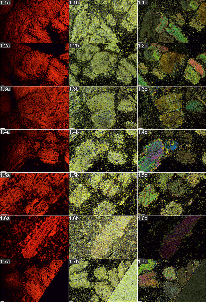
FIGURE A2. Photomicrographs of Protocinctus under cathodoluminescence (a), optical (b) and polarizing microscopy (c), respectively: 2.1-2.2, Axial to slightly oblique sections of various integument plates; 2.3, transversal section of marginal plate; 2.4, transversal to slightly oblique sections of various integument plates; 2.5, Axial to oblique section of marginal plate; 2.6, transversal section of marginal plate; 2.7, Axial to slightly oblique section of thecal? plate. Scale bar equals 100µm.
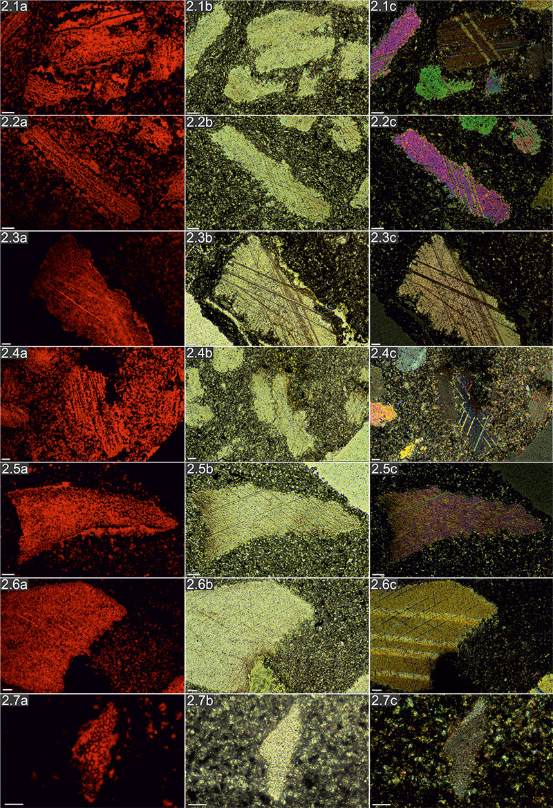
FIGURE A3. Photomicrographs of Protocinctus under cathodoluminescence (a), optical (b) and polarizing microscopy (c), respectively: 3.1-3.3, 3.5m, Axial to slightly oblique sections of various integument plates; 3.4, 3.6, Axial sections of marginal plates. Scale bar equals 100µm.
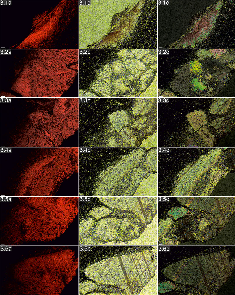
FIGURE A4. Photomicrographs of Stromatocystites under cathodoluminescence (a), optical (b) and polarizing microscopy (c), respectively: 4.1, 4.2, 4.4, 4.6, transversal to slightly oblique sections of various ambulacral plates; 4.3, 4.5, 4.7, transversal to slightly oblique sections of thecal plates. Scale bar equals 100µm.
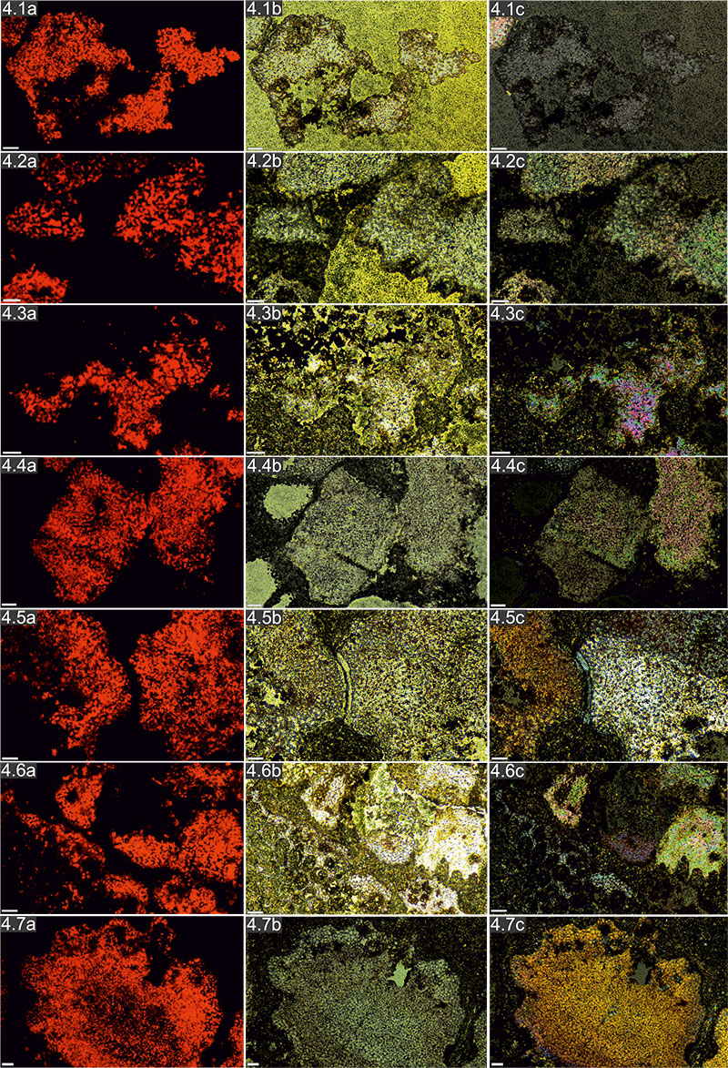
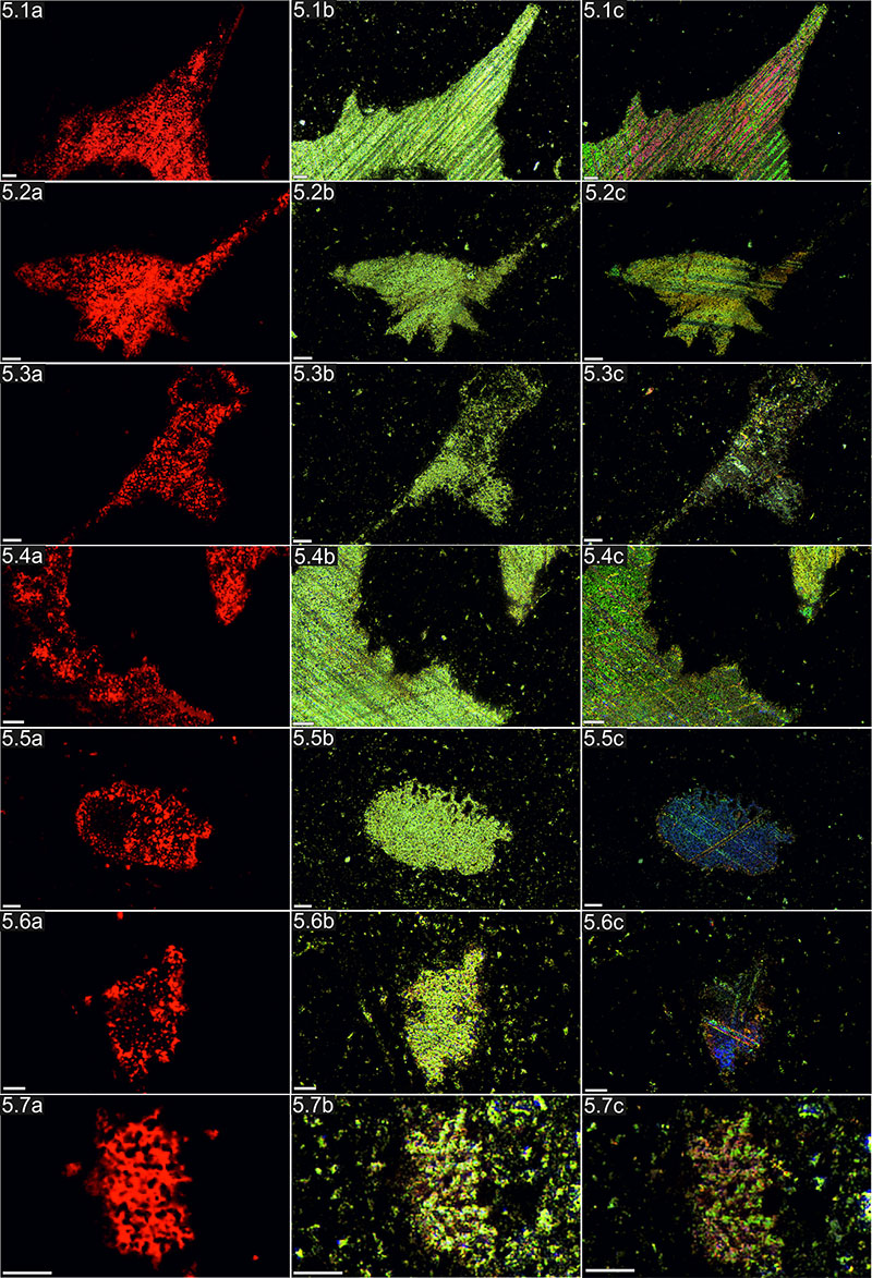
FIGURE A5. Photomicrographs of Dibrachicystidae under cathodoluminescence (a), optical (b) and polarizing microscopy (c), respectively: 5.1-5.4, longitudinal to slightly oblique sections of thecal plates; 5.5-5.7, Slightly oblique sections of columnal? plates. Scale bar equals 100µm.