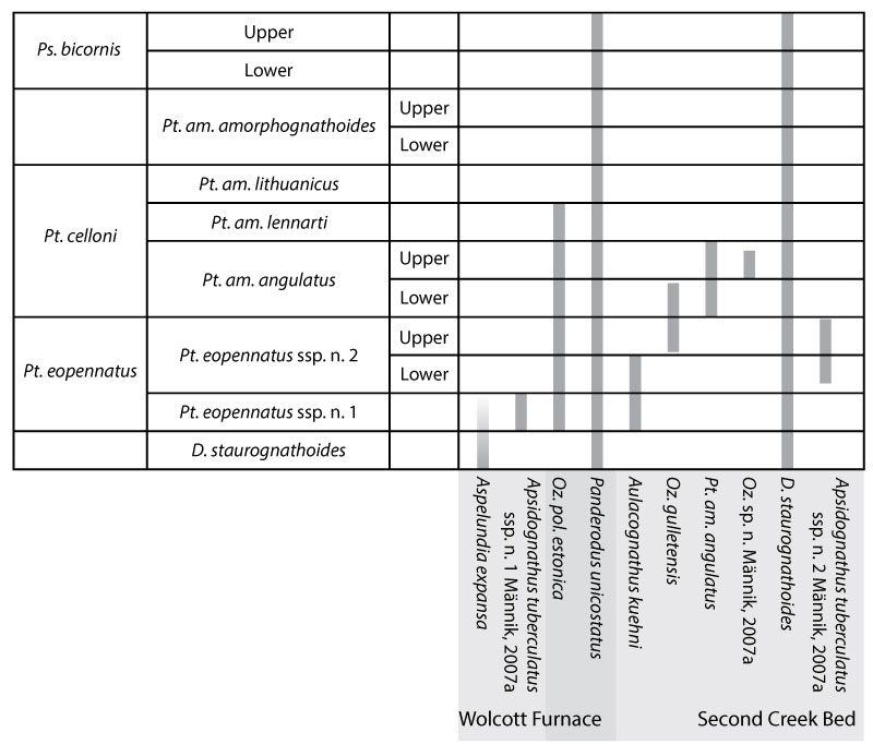
FIGURE 1. Map of the aerial extent of Silurian bedrock exposed at the surface in central and western New York State (Modified from Fisher et al., 1970). The distribution of four major lithostratigraphic divisions of the Silurian are demarcated. Monroe, Wayne, and Oneida Counties are outlined; the three localities featured in this study are indicated by dashed lines.
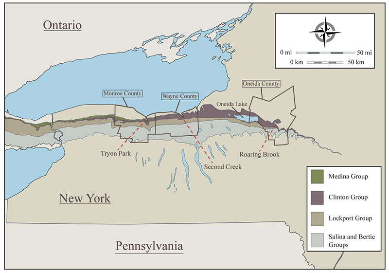
FIGURE 2. A scanned polished slab of the Second Creek Bed, collected from Second Creek. The precise level of the sample is indicated on the graphic log on the left side of the figure (key to lithologies and symbols may be found on Figure 3). The numbered boxes superimposed on the scanned image indicate the fields of three magnified views. 1. Magnified image of the upper part of a limestone rip-up clast showing the partial dissolution of the carbonate with replacement by pyrite. 2. Magnified image of phosphatized brachiopods in cross section surrounded by quartz and phosphate pebbles, with small phosphate grains sheathed by thin rinds of pyrite. 3. Upper surface of limestone rip-up clast showing a small lag of pyrite grains and a bioclast (possible crinoid) replaced by pyrite. Scale bar equals 3 cm.
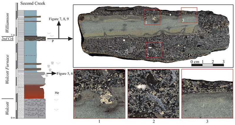
FIGURE 3. Graphic logs of outcrops exposed at the three localities featured in this study (see map on Figure 1). From left to right: Tryon Park (Monroe County), Second Creek (Wayne County), and Roaring Brook (Oneida County). Lithostratigraphic terms are shown in black italics. On the right is the sequence stratigraphic interpretation of Brett et al. (1990, 1998). The correlations of this framework are highlighted by the solid shading. Conodont biozones recognized in these successions are denoted by red lines and text (data compiled from Rexroad and Richard, 1965; Kleffner, reported in Brett et al., 1990; 1998; Loydell et al., 2007; Verniers et al., 2012; this study). Note the crossing of different correlation lines between Second Creek and Roaring Brook. A key to all lithologic symbols, labels, and patterns are shown below the correlation.
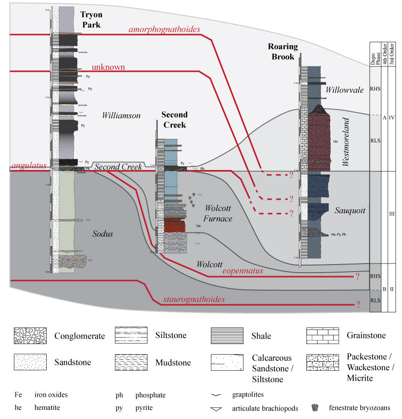
FIGURE 4. A comparison of the traditional and revised standard conodont biozonations that have been employed in the Appalachian Foreland Basin, compared to the global standard graptolite biozonation. The standard graptolite biozonation for the Telychian comes primarily from Sadler et al. (2009), Cramer et al. (2011), and Melchin et al. (2012). The modern standard conodont biozonation is based primarily on Männik (2007a) and Jeppsson (1997). The traditional standard conodont biozonation is based on the work of Walliser (1964). The correlation of these zones is derived primarily from the work of Männik (2007a), Cramer et al. (2011), and Melchin et al. (2012). The presumed age of the stratigraphic units studied herein are displayed on the right side of the figure (based on the work of Brett et al., 1998; Loydell et al., 2007; Verniers et al., 2012; this study).
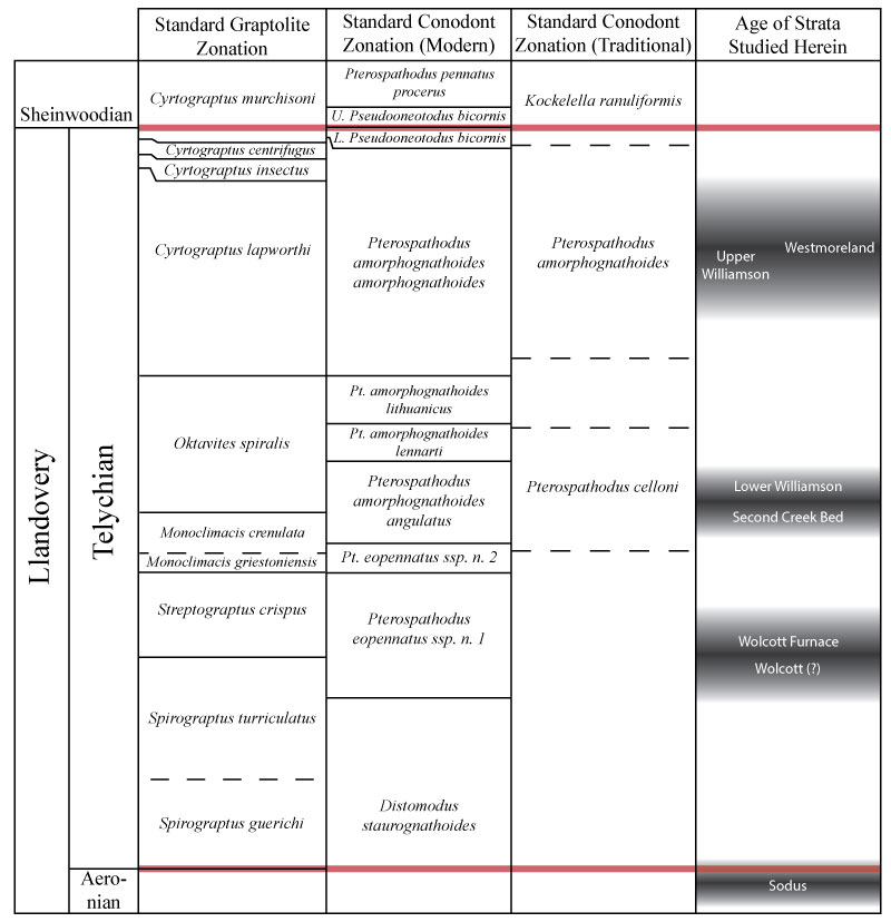
FIGURE 5. Scanning electron microscope images of selected conodonts recovered from insoluble residues of a sample of the Wolcott Furnace Hematite collected from Second Creek, Wayne County, New York (see Figure 2). 1. Aspelundia cf. A. expansa Armstrong, 1990. Posterior view of dextral Sb element, NS20110812003A. 2. Aspelundia cf. A. expansa Armstrong, 1990. Posterior view of fragmented Sa element, NS20110812003B. 3. Aspelundia cf. A. expansa Armstrong, 1990. Specimen also shows affinities toward Oulodus? australis Bischoff, 1986 or Oulodus? rectangulus sensu Bischoff, 1986. Anterior view of dextral Sb element, NS20110812003C. 4. Aspelundia cf. A. expansa Armstrong, 1990. Lateral view of Sc element, NS20110812003D. 5. Aspelundia sp. Inner lateral view of Sc element, NS20110812003E. 6. Aspelundia? sp. or Oulodus? sp. Outer lateral view of Sb element, NS20110812003F. 7.-8. Ozarkodina polinclinata estonica Männik, 1992. 7. Lateral view of fragmented Pa element, NS20110812003G. 8. Lateral view of fragmented Pa element, MK19860104A. 9. Apsidognathus tuberculatus ssp. n. 1 Männik, 2007a. Upper view of lyriform element, MK19860104B. 10. Pseudooneotodus coniformis Upper view, MK19860104C. Scale bar equals 200 μm.
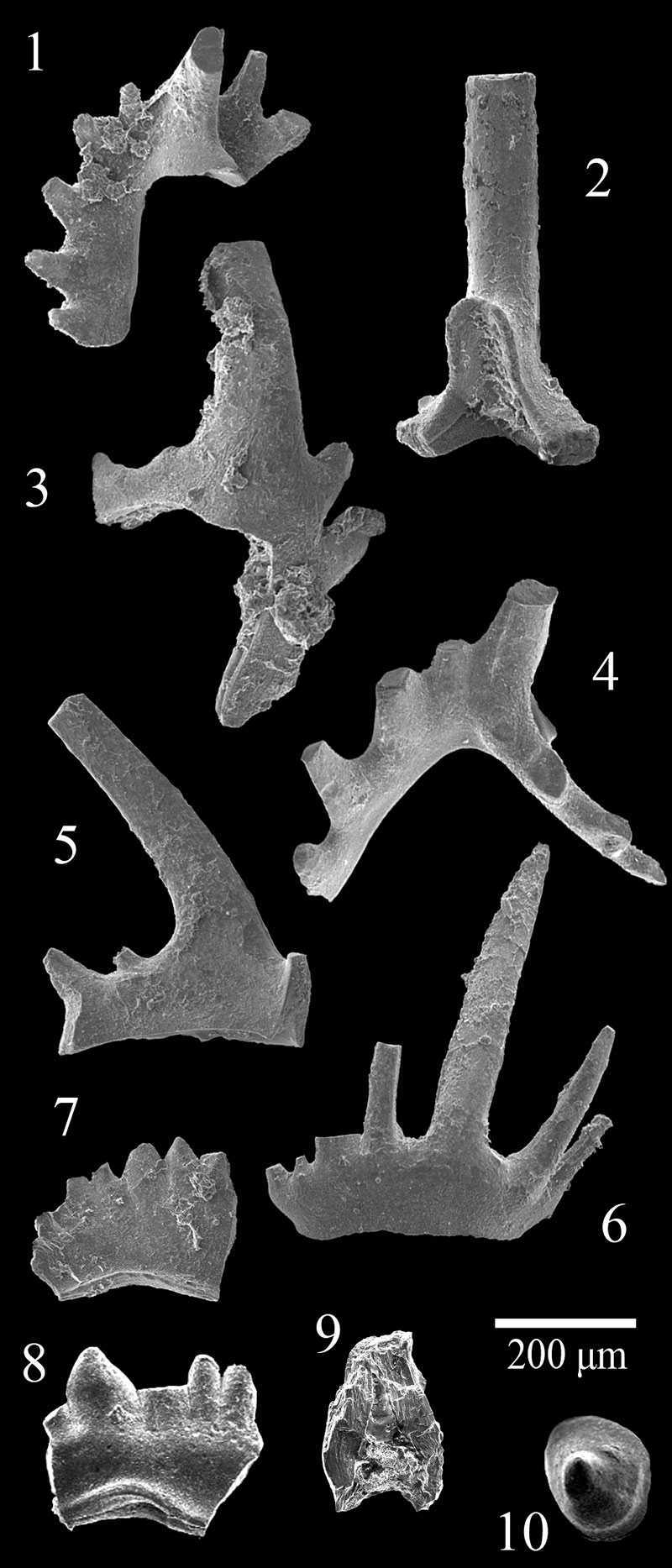
FIGURE 6. Shaded line drawings of selected conodonts recovered from insoluble residues of a sample of Wolcott Furnace Hematite collected from Second Creek, Wayne County, New York (see Figure 2). 1.-2. Ozarkodina polinclinata estonica Männik, 1992. 1. Anterior view of Sb element, NS20110812003G. 2. Posterior view of Sb element, NS20110812003G. 3.-4. Oulodus sp. or Aspelundia sp. 3. Inner lateral view of Sc element, NS20110812003H. 4. Outer lateral view of Sc element, NS20110812003H. 5. Panderodus unicostatus Branson and Mehl, 1933. Lateral view of graciliform (qg) element, NS20110812003I. 6. Panderodus unicostatus Branson and Mehl, 1933. Lateral view of arcuatiform (qa) element, NS20110812003J. Scale bar: 200 μm.

FIGURE 7. Scanning electron microscope images of conodonts recovered from insoluble residues of a sample of the Second Creek Bed collected from Second Creek, Wayne County, New York (see Figure 2). 1. Aulacognathus kuehni Mostler, 1967. Upper view of Pa element, NS20110807005A. 2. Ozarkodina polinclinata estonica Männik, 1992. Lateral view of Pa element, NS20110807005B. 3. Ozarkodina gulletensis Aldridge, 1972. Oblique upper view of Pa element, NS20110807005E. 4. Ozarkodina cf. O. sp. n. Männik, 2007a. Lateral view of Pa element, NS20110807005D. 5. Aspelundia? sp. Lateral view of Sb element, NS20110807005I. 6. Icriodella sp. Oblique upper view of Pa element, NS20110807005F. 7. Oulodus sp. Posterior view of Sb element, NS20110807005G. 8. Oulodus? petilus Nicoll and Rexroad, 1968. Lateral view of Sc element, NS20110807005H. 9. Pterospathodus sp. Lateral view of Pb element, NS20110807005C. 10.-12. Pterospathodus amorphognathoides angulatus Männik, 1998. 10. Lateral view of Pa element, NS20110807005J. 11. Lateral view of Pb1 element, NS20110807005K. 12. Lateral view of Pb1 element, NS20110807005L. 13.-16. Distomodus staurognathoides Walliser, 1964. 13. Upper view of Pa elements, NS20110807005M. 14. Upper view of Pa elements, NS20110807005N. 15. Lateral view of Pb element, NS20110807005O. 16. Lateral view of M element, NS20110807005P. 17.-19. Panderodus unicostatus Branson and Mehl, 1933. 17. Lateral view of falciform (pf) element, NS20110807005Q. 18. Lateral view of graciliform (qg) element, NS20110807005R. 19. Oblique lateral view of graciliform (qg) element, NS20110807005S. Scale bar equals 200 μm.
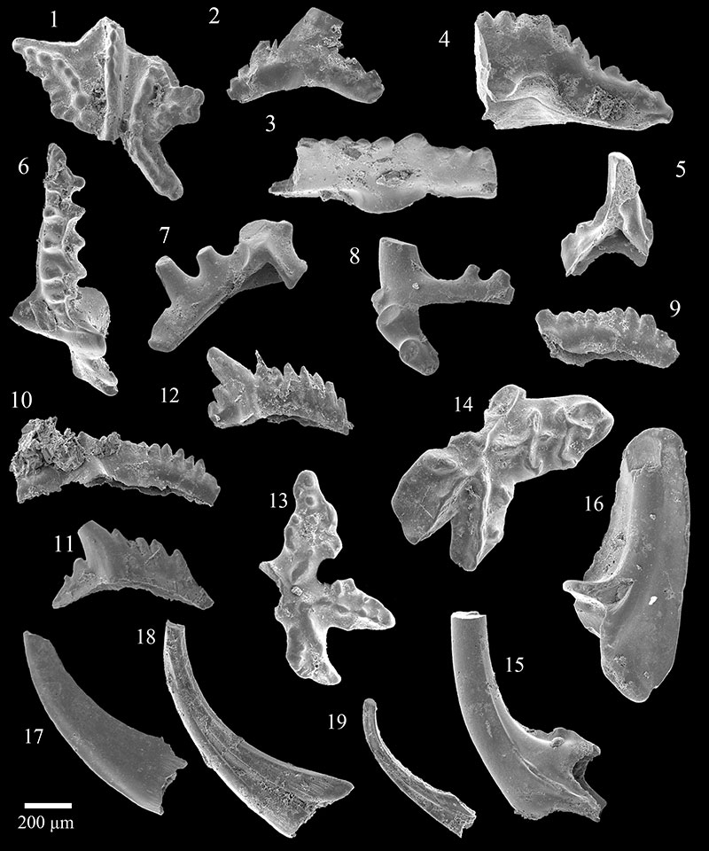
FIGURE 8. Shaded line drawings of selected conodonts recovered from insoluble residues of a sample of the Second Creek Bed collected from Second Creek, Wayne County, New York (see Figure 2). 1. Ozarkodina sp. n. Männik, 2007a. Lateral view of Pa element, NS20110807005S. 2. Apsidognahus tuberculatus ssp. n. 2 Männik, 2007a. Upper view of lyriform element, NS20110807005T. 3. Icriodella sp. Lateral view of Pa element, NS20110807005U. 4. Distomodus staurognathoides Walliser, 1964. Upper view of Pa element, NS20110807005V. 5.-6. Pterospathodus amorphognathoides angulatus Männik, 1998. 5. Lateral view of Pa element, NS20110807005W. 6. Upper view of Pa element, NS20110807005W. Scale bar equals 200 μm.
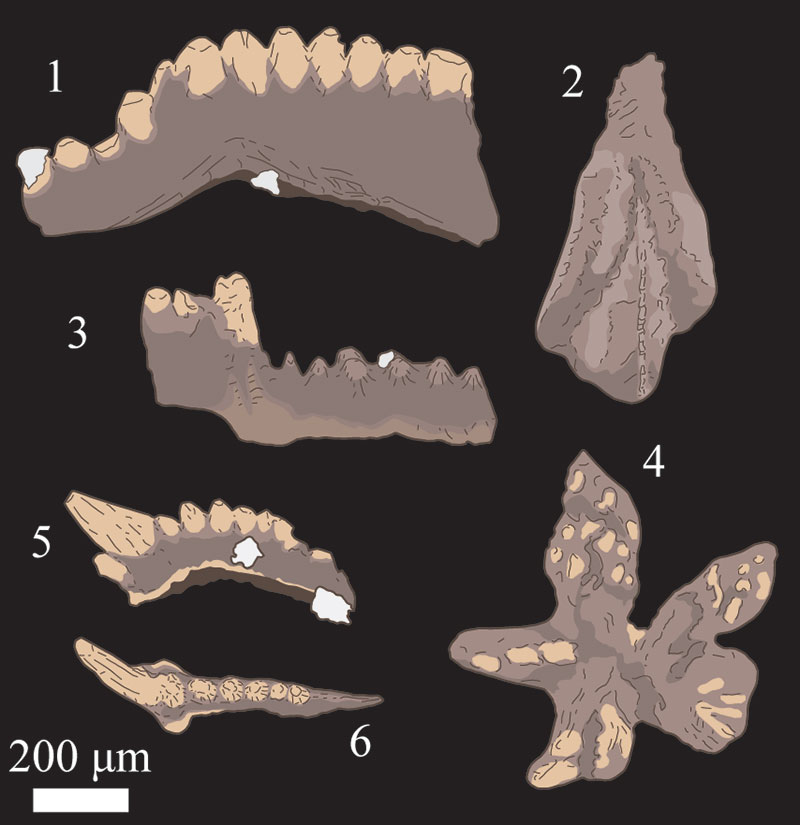
FIGURE 9. Scanning electron microscope images of the pyritized internal molds of mollusks collected from the insoluble residues of a sample of the Second Creek Bed, collected from Second Creek, Wayne County, New York (see Figure 2). 1.-2. Gastropods. 3.-5. Bivalves. 6.-8. Tentaculitids. Scale bar equals 200 μm.
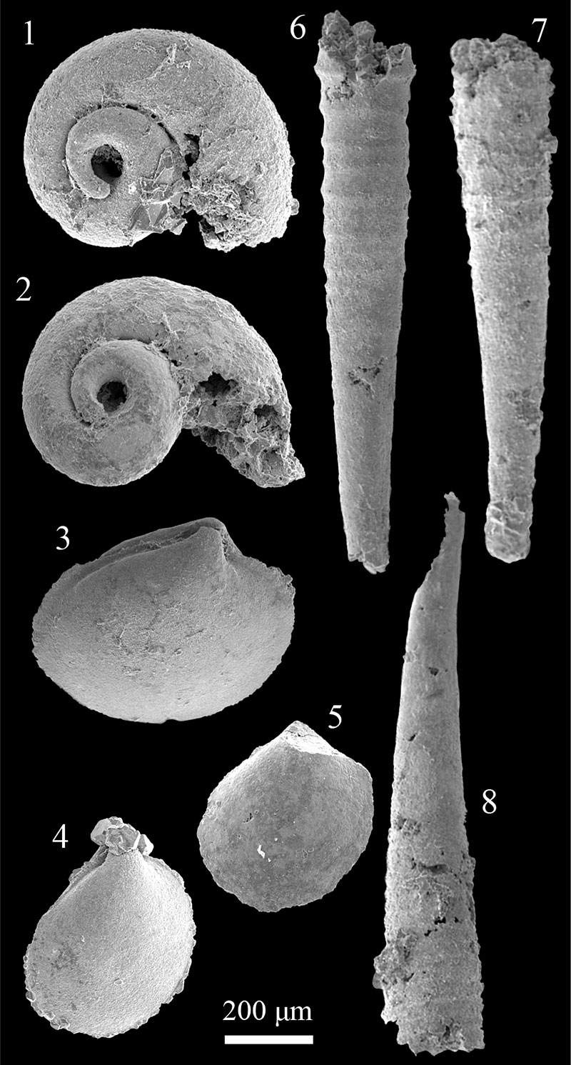
FIGURE 10. Polished sample of the rippled conglomeratic horizon collected 70 centimeters below the base of the Westmoreland Hematite at Roaring Brook. Numbered boxes superimposed on the scanned polished slab indicate the fields of three magnified views. 1. View of quartz and phosphate pebbles surrounded by smaller, sand sized grains, many of which are composed of, or coated with a dark red mineral of unknown composition. 2. Black phosphate pebble partially coated by dark red minerals and iron sulfides. 3. Quartz and phosphate pebbles interspersed among sand sized grains of a red mineral of unknown composition. Many of which are coated by a light grey to pink minerals, also of uncertain composition. Scale bar equals 3 cm.
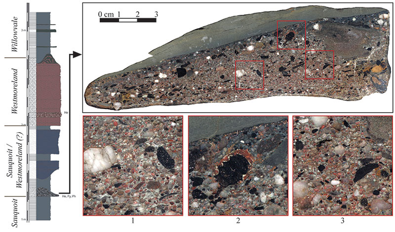
FIGURE 11. Known ranges of conodont taxa recovered from the Wolcott Furnace Hematite and Second Creek Bed (modified from Männik, 2007a, b).