FIGURE 1. CT slices and digital reconstruction of the skull of Teyumbaita sulcognathus. (1) Digital reconstruction of the skull with the slicing planes showed in (2), (3), and (4). (2) Coronal slice of the top of the skull with the impressions of the olfactory portion of the brain. The dashed line marks the location of a shallow keel under the frontals which separates the olfactory bulbs. (3) Sagital slice with the preserved portion of the endocast. (4) Axial slice of the snout. Scale bar equals 5 cm, in (2), (3) and (4). Abbreviations: A, anterior; B, bottom; d.e.v, dorsal expansion of the vomer; d.p, dorsal peak of the endocast; e, endocast; L, left; n.c, nasal cavity; o.b, olfactory bulb; o.t, olfactory tract; P, posterior; R, right; T, top.
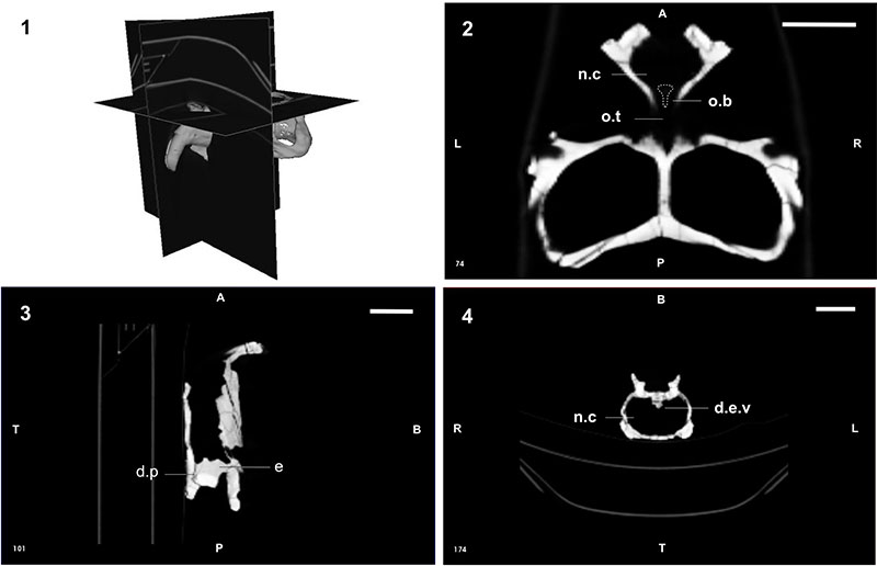
FIGURE 2. In (1) skull of Teyumbaita sulcognathus digitally rendered showing the position of a coronal slice (2), in which is possible to note a cast of what once was interpreted by Benton (1983) as the cast of a pineal organ in Hyperodapedon gordoni. However, the present data suggest that this structure is actually part of the dorsal peak of the endocast of T. sulcognathus and it represents probably a venous sinous or a dural expansion. Scale bar equals 5 cm, in (2). Abbreviation: d.p, dorsal peak.
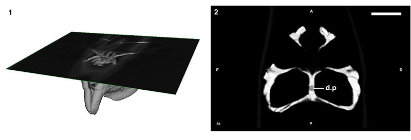
FIGURE 3. Cranial endocast of Teyumbaita sulcognathus. (1) Semi-transparent skull for allowing the visualization of the endocast. The dotted line represents the inferred anterior contours of the brain based on the impressions of the olfactory bulbs. (2) Cranial endocast in left lateral view. Scale bars equal 5 cm and 1 cm in (1) and (2), respectively. Abbreviations: c, cerebellum; c.h, cerebral hemispheres; d.p, dorsal peak of the endocast; e.n, external nares; i.e, left inner ear; m.f, metotic foramen; o, orbit; o.b, olfactory bulb; o.t, olfactory tract.
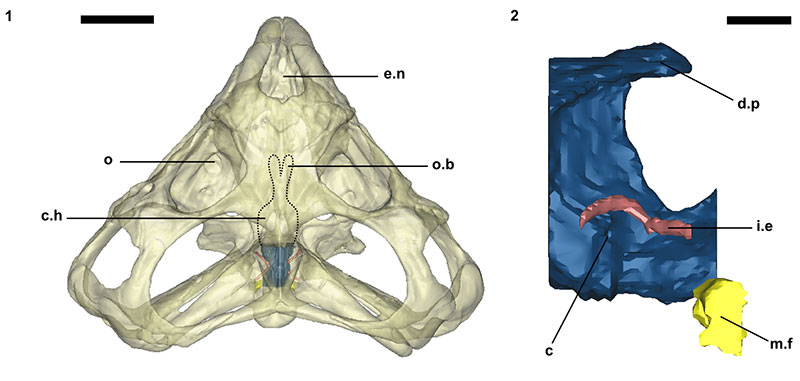
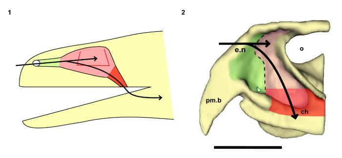
FIGURE 4. Nasal cavity of a generalized tetrapod (1) and the digital reconstruction of the right half of the snout of Teyumbaita sulcognathus in medial view (2). The horizontal and vertical arrows represent the airstream passing, respectively, to the olfactory part of the nasal cavity and the oral cavity and then the upper respiratory tract. In green, the nasal vestibule, which is followed by the nasal cavity artificially colored in pink. A concha is located within the nasal cavity proper of the generalized tetrapod and has its contours in red. The last portion of the nasal capsule is the nasopharyngeal duct here indicated in red, but its presence in T. sulcognathus is speculative. The dashed line indicates the course of the internal ridge that possibly marks the boundary between the vestibule anteriorly and the nasal cavity proper posteriorly. Scale bar equals 5 cm, in (2). Abbreviations: ch, choana; e.n, external nares; o, eye orbit; pm.b, pre-maxillary beak.