FIGURE 1. Giant deer skull. 1. frontal view. 2. back view of the boney outgrowth with scanned plane.
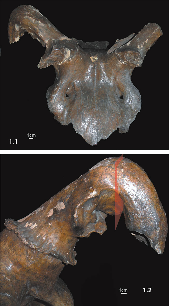
FIGURE 2. 1. Radiography of giant deer antler (explanations see text). 2. CT images of giant deer antler (explanations see text).
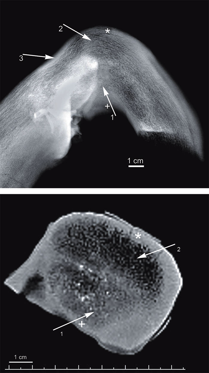
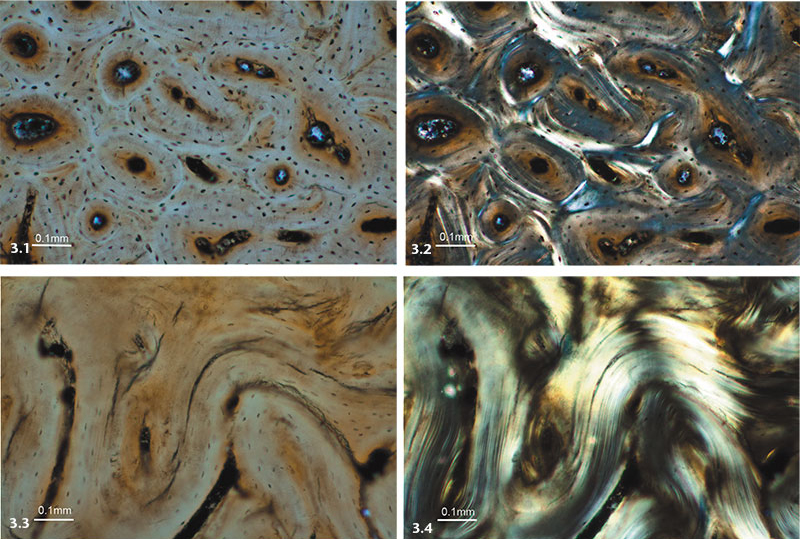
FIGURE 3. 1. Histological image of giant deer antler, normal tissue in white light (explanations see text). 2. Same fragment in polarised light (explanations see text). 3. Tissue of abnormal structure in white light (explanations see text). 4. Same fragment in polarized light (explanations see text).