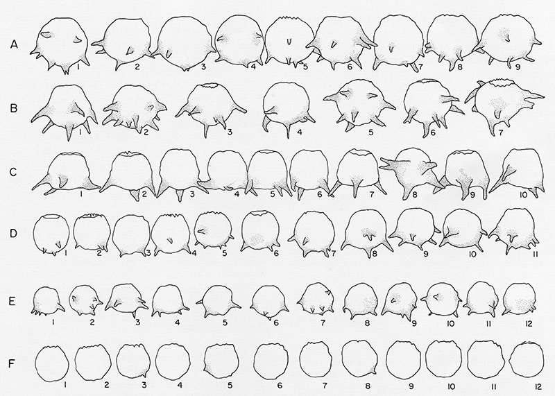
FIGURE 1. Specimens of Difflugia protaeiformis Lamark, 1816, the type species of Difflugia. 1. Difflugia protaeiformis Lamarck, 1816 as illustrated in Lamarck, 1816, pl. 17, figure. 5. 2. and 3. Scanning electron micrographs of hypotypes of Difflugia protaeiformis Lamark strain “protaeiformis” from Mytopo Lake, eastern Ontario (Latitude 44°40.179N, Longitude 77°3.397W). Scale bar equals 100 μm. 2. side view of typical specimen with characteristic broad base and narrow, elongate neck; 3. side view of a relatively common morphology characterized by heavy agglutination on neck.
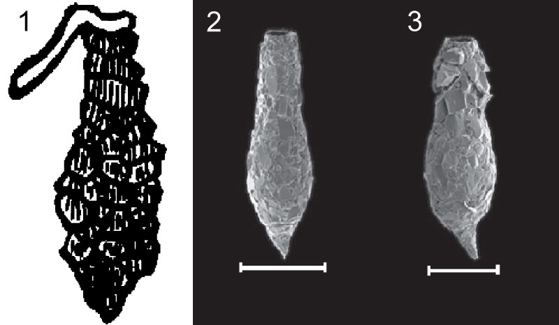
FIGURE 2. Specimens of Mediolus corona from Wallich, 1864.1. illustrated as figure 19 in the original publication, Wallich described the specimen as “front view, showing crenulate margin of aperture; a six-horned variety”; 2. illustrated as figure 20 in the original publication, Wallich described the specimen as “a somewhat smaller four-horned variety; side view.”
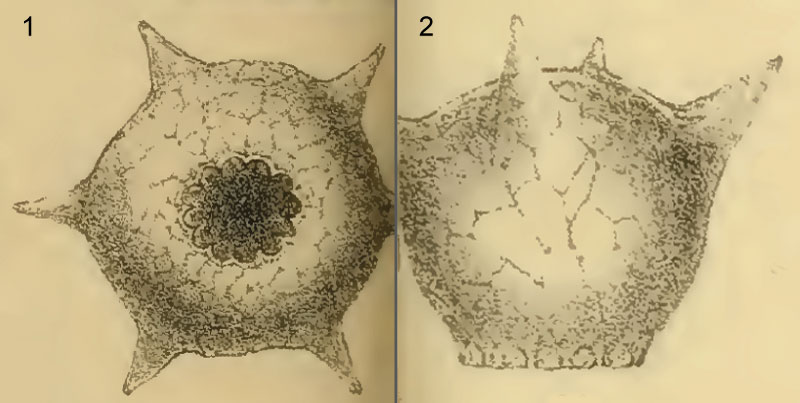
FIGURE 3. Specimens of Mediolus corona from lakes in the Subarctic (“Lake 10” near Yellowknife, Northwest Territories; 62°30.924’N, 113°46.817’W); temperature region (Mew Lake, Algonquin Park, Ontario; 45°34.575’N, 78°31.087’W, Bell’s Lake, King (Greater Toronto Area), Ontario; 43°N56.597, 79°39.762W); and equatorial regions (Holocene core from Laguna de Quistococha, Iquitos, Peruvian Amazon; 3°49.746’N, 73°19.157’W). 1. side view of typical spheroidal hypotype (CANA 91905) from sediment-water interface in Mew Lake showing orientation of basal spines and thin narrow apertural rim; 2. oblique apertural view of same specimen showing crenulated aperture; 3, apertural view of hypotype (CANA 87197) from sediment-water interface in Bell’s Lake showing round aperture; 4. Closeup of individual crenulation in Bell’s Lake specimen showing spiny projections; 5. Side view of globular hypotype (CANA 87186) from “Lake 10” showing distribution of spines; 6. oblique apertural view of same specimen showing narrow apertural rim; 7. apertural view of same specimen showing rounded apertural opening and regular distribution of basal spines; 8. Gobular hypotype (CANA 87198) from Holocene core collected from Laguna de Quistococha with relatively few short spines; 9. apertural view of different hypotype (CANA 87199) from Laguna de Quistococha showing typical rounded and crenulated aperture but with considerably reduced basal spines than shown in “Lake 10” specimen (7 above); 10. oblique apertural view of same specimen showing typical narrow crenulated apertural lip; 11. close up of apertural region on same specimen showing regular spacing and uniform structure of crenulations; 12. close up of a single crenulation showing typical spiney ultrastructure, which are nearly identical to those of temperature latitude specimens (4 above).
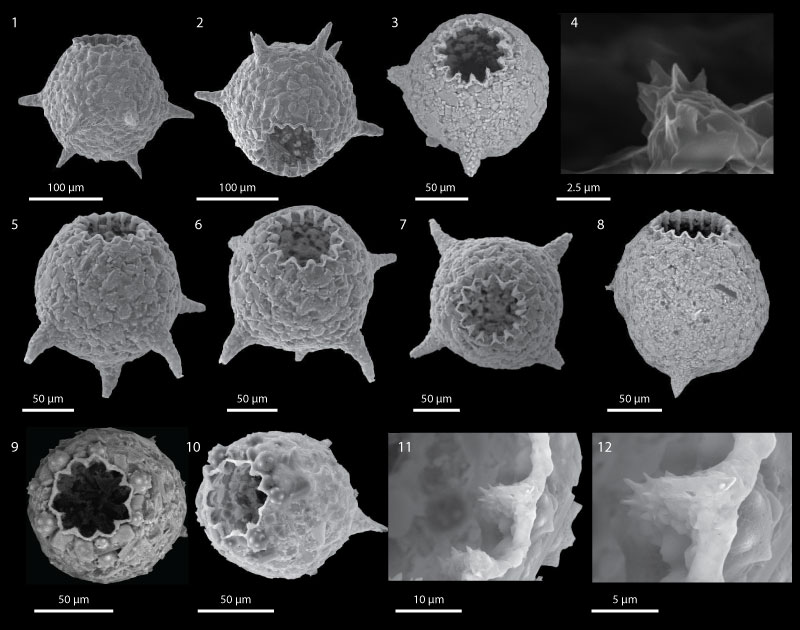
FIGURE 4. Line drawings of Mediolus corona clones produced within six of Jennings (1916) broods, redrafted and reaarranged by Medioli and Scott (1983). The figure shows that although there is little variation within broods through only a few generations there is considerable variation in both size and process number and arrangement between broods. The research of Jennings (1916) indicated that although the crenulated aperture was a constant feature in all clones, some morphotypes can be processless.