FIGURE 1. 1.Simplified geological scheme for the study area and location of fossil sites (modified after Baldanza et al., 2011). 2. Bargiano and Montemoro sedimentological and biostratigraphic sections. Pictures (m1, m2) show the emergence of large cololites in the Montemoro section. Grain-size scale: C = Clay, S = Silt, VFS = Very Fine Sand, FS = Fine Sand. bmG= base of medium Gephyrocapsa event; blG= base of large Gephyrocapsa event (sensu Raffi, 2002).
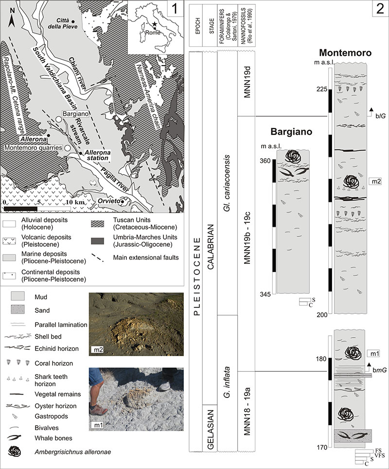
FIGURE 2. Bargiano ichnofossils-bearing site. 1., 3. Panoramic views of cololites in W-E (1) and N-S ( 3) directions. 2. Simplified topographic map with localization of cololites and whale remains; main bed’s attitude is reported. 4-6. Details of whale bones. 4. Part of the skull, associated to shell beds. 5. Cervical fused vertebrae 2nd-7th (Ve). 6. Oyster shells (Oy) grown on a bone surface.
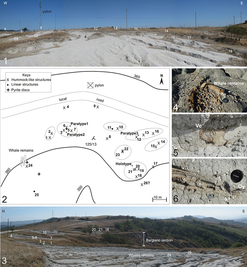
FIGURE 3.Simpler cololites (intestinelite group) from Bargiano section. 1., 7. Typical irregular elongated mass (CT02), produced by different helicoidal swirls, Bargiano section, in lateral view (1) and backside view ( 7). Arrows indicate the occurrence of longitudinal striae. 2. Mass (CT03) with bulges (rognons). 3. Holotype of Ambergrisichnus alleronae(CT01), showing converging striae in the left apex (arrow) and enlargement at the other apex. 4. Characteristic helicoidal arrangement and rings with different colours (CT04). 5-6. Side view (5) and front view (6) of rings (CT04), (modified after Baldanza et al., 2013). 8-9. Modern examples of ambergris masses with concentric rings and changes in colour, comparable to study specimens (arrow in 8 indicates a squid beak).
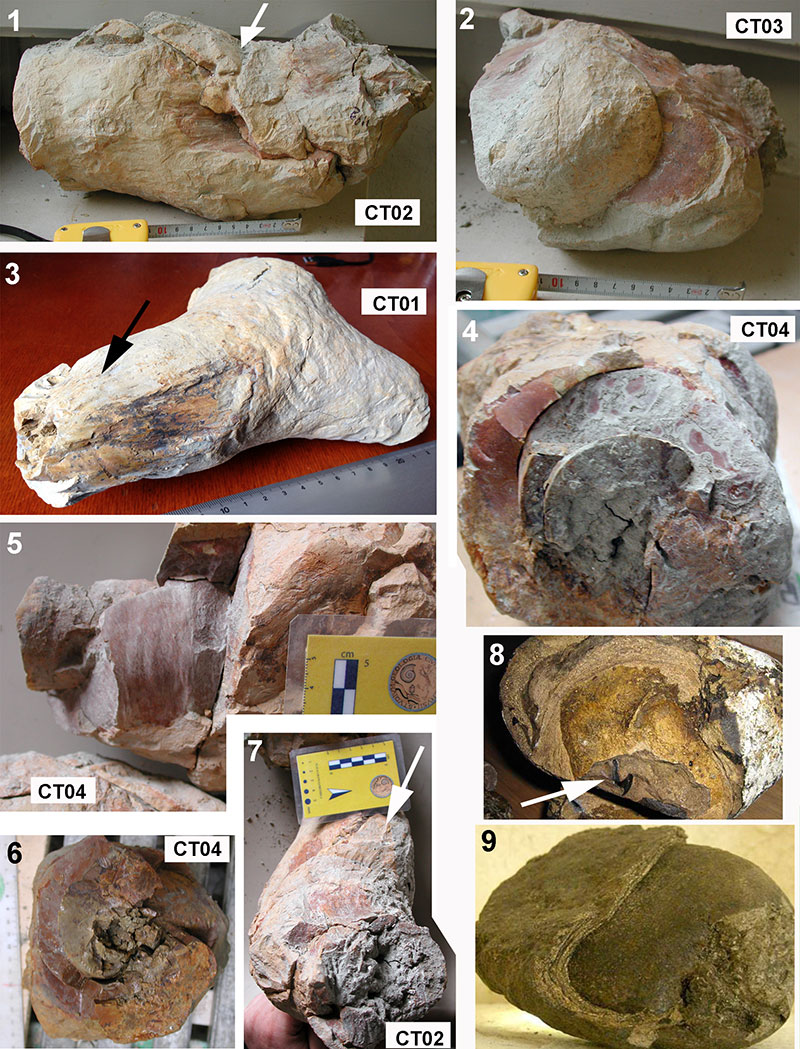
FIGURE 4.Complex cololites (intestinelite group), Bargiano section. 1., 4. Hummock-like cololite (n 8 in Figure 2.2), 50 cm high and 80 cm wide, with a concave, irregular center and outer sub-circular to elliptical tunnels ( 1). Disaggregated portions ( 4) reveal both striae and concentric structure. 2. Hummock-like cololite (n 12 in Figure 2.2) formed by an accumulation of irregular, elongated slabs. 3. Complex hummock-like structure (n 15 in Figure 2.2) with irregular, meandering tunnels disposed at various levels. 5. Isolated linear cololite (n 25 in Figure 2.2), 20 cm in diameter, partially emerging from clay deposits. 6. Irregular, 60 cm long linear cololite (n 11 in Figure 2.2), showing spiral coiling at the right side.
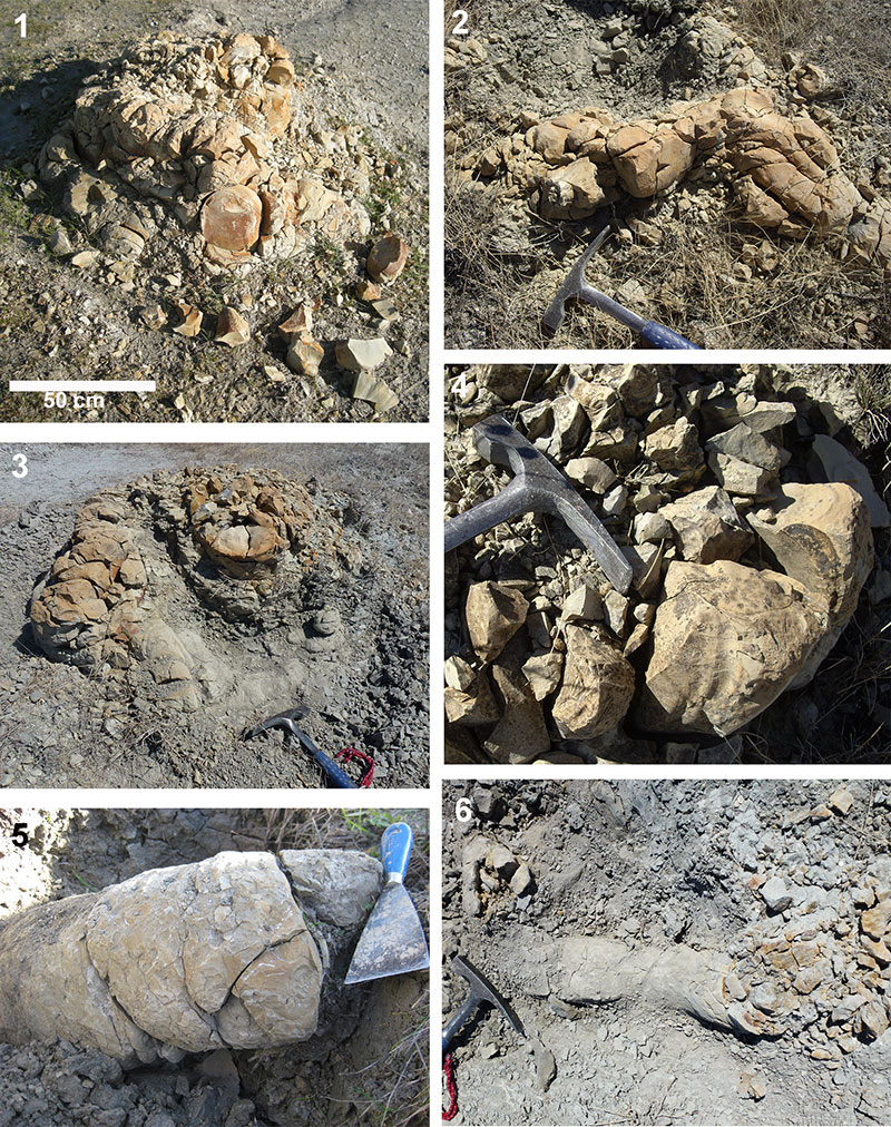
FIGURE 5. Squid beaks from structure n 8 (Figure 2.2). Part of a lower beak ( 1) and longitudinal section of a beak ( 2), emerging from the rough surface of rock sample. 3-4. Microscopic features of squid beaks inside cololites (structure n 12,Figure 2.2). Crystals and framboids of pyrite ( 4), scattered into the micropeloidal matrix with dolomite microcrystals are also visible (1 and 4 are modified after Baldanza et al., 2013).
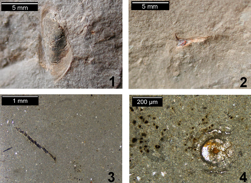
FIGURE 6.XRPD patterns ( 1) and mass changes in thermogravimetric analysis ( 2) of five cololite samples. Numbers of samples correspond to the structure of provenance (Figure 2.2).
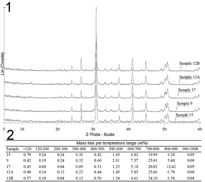
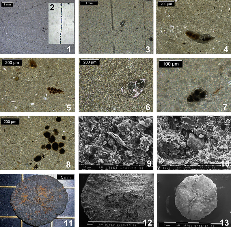
FIGURE 7. 1, 3-8.Transmitted light photomicrographs of petrographic thin sections of the Bargiano cololites. 1. Interior of cololite (structure n 12) showing a micropeloidal matrix crossed by long chains of pyrite microgranules. 2. Beggiatoa specimens, sulphur large bacteria, single free-living filament with sulphur inclusion. 3. Detail of fossil Beggiatoa-like filaments from structure n 12. 4. Subspherical, red-brownish micromasses of pyrite (or sulphur), surrounded by a grey “halo” that inglobates all. To note the micropeloidal matrix. This structure may be comparable with large Thioploca cells. 5-7. Benthic foraminifera, partially and/or totally filled by bacteria-induced microcrystals of pyrite. 8. Pyrite framboids from structure n. 12. 9-10. SEM images of cololite internal fragments.The both surfaces are fresh, no acid attack was made. 9. Rosette structures made of dolomite microcrystals, and scattered single spherical cells referable to bacteria. In the centre, mica crystals with typical shape. 10. Dolomite crystals with well-developed orthorhombic shape (at central right), sparse spherical cells of bacteria, and a quartz crystal. Dolomite crystals (left) are aggregated to form a compact mass. 11-13. Bacteria-induced pyritization (sun-like pyrite discs). 11. Stereomicrophotograph of a large pyrite disc (about 1.5 cm in diameter). 12. SEM image of disc surface (particular), showing a large amount of spherical cells, progressively smaller form center to periphery. This arrangement is associated to bacteria colonies. 13. Small pyrite disc, yellow/orange, with elliptical to dumb bell shaped bacteria bodies arranged in a short chain.